. 2003 Sep 2;100(18):10477-82.
doi: 10.1073/pnas.1832245100. Epub 2003 Aug 19.

Chaperone-mediated in vitro assembly of Polyomavirus capsids

Affiliations

Chaperone-mediated in vitro assembly of Polyomavirus capsids

Laura R Chromy et al. Proc Natl Acad Sci U S A. .

Abstract

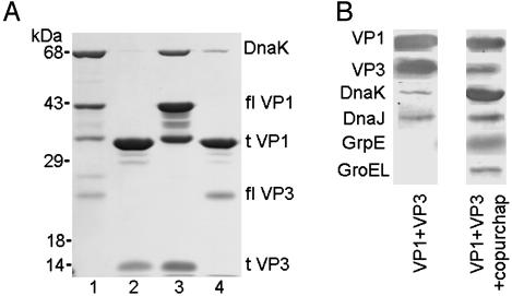
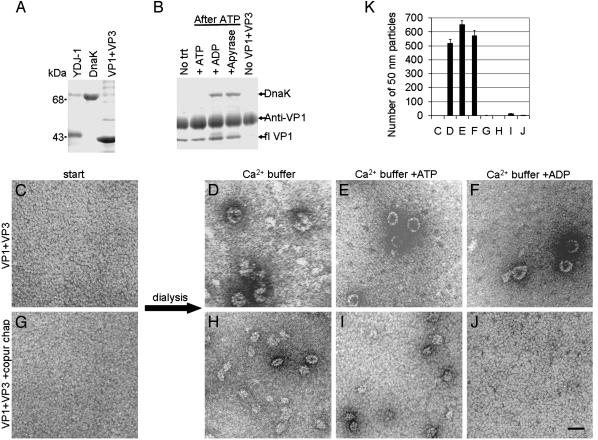
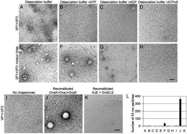
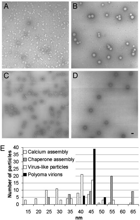
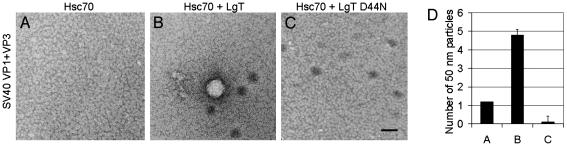
The polyomavirus coat protein viral protein 1 (VP1) has the intrinsic ability to self-assemble in vitro into polymorphic capsid-like structures on addition of calcium. In contrast, polyomavirus assembly in vivo is rigorously controlled, such that virions of uniform size are formed only in the cell nucleus. During viral infection, the 72 kDa cellular chaperone heat shock cognate protein (hsc70) binds VP1 posttranslation and colocalizes with VP1 to the nucleus, thereby suggesting a role for approximately 70-kDa heat shock protein (hsp70) family chaperones in regulating the quality and location of capsid assembly. We found that, after expression of recombinant VP1 in Escherichia coli, the prokaryotic hsp70 chaperone DnaK copurified with the VP1 C-terminal domain that links pentamers in an assembled capsid. When stably bound to VP1, DnaK inhibited in vitro assembly induced by calcium. However, in the presence of ATP, the hsp70 chaperone system comprised of DnaK, DnaJ, and GrpE assembled VP1 into uniform capsids without requiring calcium. Chaperone-mediated assembly was similarly catalyzed by the eukaryotic hsc70 protein, in combination with the J-domain function of the simian virus 40 large T-antigen protein. Thus, polyomavirus capsid assembly can be recapitulated with high-fidelity in vitro using either prokaryotic or eukaryotic hsp70 chaperone systems, thereby supporting a role for cellular chaperones in the in vivo regulation of virion assembly.


Figures

Fig. 1.
Fig. 1.
E. coli chaperones interact with the C-terminal domain of the polyomavirus capsid protein VP1. (A) Recombinant VP1 and a GST-VP3 fusion protein were coexpressed in E. coli and were purified by using glutathione Sepharose chromatography, followed by thrombin cleavage as described (15). Eluates were analyzed by SDS/PAGE and Coomassie blue staining. Lane 1, full-length VP1 (flVP1) coexpressed with full-length VP3 (flVP3); lane 2, residues 32-316 of VP1 (tVP1) coexpressed with the C-terminal 105 residues of VP3 (tVP3); lane 3, flVP1 coexpressed with tVP3; and lane 4, tVP1 coexpressed with flVP3. (B) Immunoblots of VP1 + VP3 pentamers purified by glutathione affinity, ion exchange, and gel filtration chromatography (VP1 + VP3), or only glutathione affinity and gel filtration chromatography (VP1 + VP3 + copurified chaperones).
Fig. 2.
Fig. 2.
A stable interaction between E. coli chaperones and VP1 inhibits calcium-mediated assembly. (A) Purified proteins YDJ-1, DnaK, and VP1 + VP3 analyzed by SDS/PAGE and Coomassie blue staining. (B) Anti-VP1 (I58) (7, 22) coimmunoprecipitation of purified proteins (YDJ-1 at 0.5× molar concentration, DnaK at 4× molar concentration, and VP1 + VP3 at 1× molar concentration) incubated without treatment (No trt), which is incubated briefly in ATP, followed by either ATP, excess ADP, or apyrase; or incubation of YDJ-1 and DnaK in the absence of VP1 + VP3 (No VP1 + VP3). (C-G) Purified pentamers of recombinant VP1 + VP3 without chaperones (C) or with copurified E. coli chaperones (G), in dissociating buffer visualized by negative stain and TEM. (D-F and H-J) In vitro assembly reactions of VP1 + VP3 pentamers without (D-F) or with (H-J) copurified chaperones, after dialysis into indicated calcium buffers, and visualized by negative stain and TEM. (Scale bar, 50 nm.) (K) Quantitation of the mean number of 50-nm particles per grid square (±SEM, n = 3) from the in vitro assembly reactions shown in C-J.
Fig. 3.
Fig. 3.
E. coli chaperones DnaK, DnaJ, and GrpE assemble capsid proteins in vitro. (A-H) In vitro assembly reactions of VP1 + VP3 pentamers without (A-D) or with (E-H) copurified chaperones dialyzed into indicated dissociation buffers and visualized by negative stain and TEM. (I-K) In vitro assembly reactions of VP1 + VP3 pentamers at 1× molar concentration in dissociating buffer with ATP, using no chaperones (I), a reconstituted chaperone system with purified DnaK (0.5×), DnaJ (0.05×), and GrpE (0.05×)(J), or a reconstituted chaperone system with purified DnaK (0.5×), DnaJ (0.05×), GrpE (0.05×), and GroELS (1×) visualized by negative stain and TEM. (Scale bar, 50 nm.) (L) Quantitation of the mean number of 55-nm particles per grid square (±SEM, n = 3) from the in vitro assembly reactions shown in A-K.
Fig. 4.
Fig. 4.
Capsids assembled by chaperones in vitro are uniform. (A-D) Negative stain and TEM of calcium assembly reaction (A), chaperone assembly reaction (B), VP1 VLPs (C), and polyoma virions (D). (Scale bar, 50 nm.) (E) Size polymorphism, based on 50 random assembled particles for the reactions shown in A-D.
Fig. 5.
Fig. 5.
Mammalian hsc70 requires the J-domain of large T antigen to assemble capsid proteins in vitro.(A-C) In vitro assembly reactions of purified recombinant SV40 capsid proteins VP1 + VP3 at 1× molar concentration with mammalian hsc70 (1×) (A), mammalian hsc70 (1×) + LgT (at 0.1× for the J-domain) (B), or mammalian hsc70 (1×) + LgT D44N, a J-domain mutant (0.1×)(C), visualized by negative stain and TEM. (Scale bar, 50 nm.) (D) Quantitation of the mean number of 50-nm particles per grid square (±SEM, n = 10) from the in vitro assembly reactions shown in A-C.

Similar articles

Cited by

References

    1. Rayment, I., Baker, T. S., Caspar, D. L. & Murakami, W. T. (1982) Nature 295, 110-115. - PMC - PubMed
    1. Liddington, R. C., Yan, Y., Moulai, J., Sahli, R., Benjamin, T. L. & Harrison, S. C. (1991) Nature 354, 278-284. - PubMed
    1. Griffith, J. P., Griffith, D. L., Rayment, I., Murakami, W. T. & Caspar, D. L. (1992) Nature 355, 652-654. - PubMed
    1. Garcea, R. L., Salunke, D. M. & Caspar, D. L. (1987) Nature 329, 86-87. - PubMed
    1. Salunke, D. M., Caspar, D. L. & Garcea, R. L. (1989) Biophys. J. 56, 887-900. - PMC - PubMed

Publication types

MeSH terms