. 2003 Sep;133(3):334-43.
doi: 10.1046/j.1365-2249.2003.02226.x.

CD8beta/CD28 expression defines functionally distinct populations of peripheral blood T lymphocytes

Affiliations

CD8beta/CD28 expression defines functionally distinct populations of peripheral blood T lymphocytes

S Werwitzke et al. Clin Exp Immunol. 2003 Sep.

Abstract

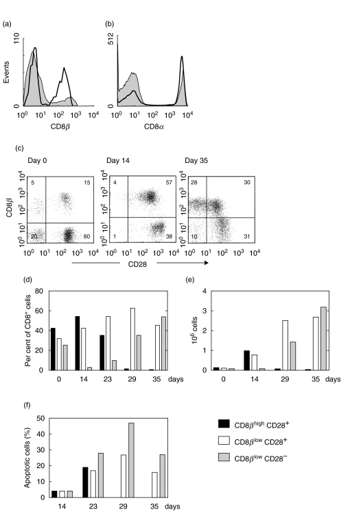
Peripheral blood CD8+ T lymphocytes generally express the CD8 coreceptor as an alphabeta heterodimer. On these cells, the CD8beta chain is present either at high (CD8betahigh) or low density (CD8betalow). CD8betahigh cells are CD28+, whereas CD8betalow cells are CD28+ or CD28-. Therefore, three subpopulations of CD8+ T cells can be described: (i) CD8betahighCD28+ (ii) CD8betalowCD28+, and (iii) CD8betalowCD28- cells. Phenotypic and functional characterization of these CD8+ T cell subsets revealed significant differences. CD8betahighCD28+ cells predominantly express CD45RA. In contrast, CD8betalowCD28+ cells frequently express CD45R0 and the activating NK receptor CD161. CD8betalowCD28- cells frequently revert to the CD45RA phenotype. In addition, these cells express CD16, CD56, CD94, and the killer-inhibitory receptors NKB1 and CD158a. Intracellular IL-2 was frequently detected in CD8betahighCD28+ cells and CD8betalowCD28+ cells, but not CD8betalowCD28- cells. CD8betalowCD28+ cells and CD8betalowCD28- cells frequently stained positive for IFN-gamma. In addition, these cells contain intracellular perforin and granzyme A. Expression of Fas (CD95) as well as susceptibility to apoptosis is markedly increased in CD8betalowCD28+ and CD8betalowCD28- cells as compared to CD8betahighCD28+ cells. In vitro activation of peripheral blood lymphocytes triggered expansion of CD8betahighCD28+ cells as well as a development into CD8betalowCD28+ and CD8betalowCD28- cells. Similarly, activation of CD8betahighCD28+ cord blood cells resulted in the appearance of CD8betalowCD28+ and CD8betalowCD28- cells. These data suggest that CD8betahighCD28+ cells can differentiate into CD8betalowCD28+ and CD8betalowCD28- cells upon TCR stimulation. Therefore, the CD8beta/CD28 subsets in peripheral blood may reflect distinct stages of post-thymic CD8+T cell development.


Figures

Fig. 1
Fig. 1
CD8 expression on peripheral blood lymphocytes from a representative healthy young donor. (a) CD8α on CD3+ gated PBL. (b) D8β on CD3+ gated PBL. CD8β is expressed either on a high level (CD8βhigh, marker 1) or on a low level (CD8βlow, marker 2). (c) –(f) PBL gated based on FSC/SSC. Percentages in respective quadrants and regions are indicated. (c) CD8βhigh cells (R1) and CD8βlow cells (R2) express comparable amounts of CD8α. (d) CD8β staining intensity is equal for MoAbs 2ST8·5H7 and 5F2 indicating that all CD8β is present in αβ heterodimers. (e) CD8βhigh cells (R1) and CD8βlow cells (R2) are CD3 positive. (f) Coexpression of CD8β and CD28 defines three populations: CD8βhighCD28+ (R1), CD8βlowCD28+ (R2), CD8βlowCD28 (R3). (g) Frequency of the three CD8β/CD28 populations in 49 healthy subjects aged 23–80 years (given as per cent of CD8β+ cells; horizontal lines indicate the median).
Fig. 2
Fig. 2
Expression of characteristic cell surface molecules on CD8βhighCD28+ (red), CD8βlowCD28+ (green), and CD8βlowCD28 cells (blue). Scatter plots are representative examples from the same healthy donor. Percentages in respective quadrants and regions are indicated. (a)Gated for PBL based on FSC/SSC. (b)– (i)Gated for CD8β+ cells. Bar charts under the plots show the median percentage ± SD of cells expressing the indicated marker in a number of individuals (n). Significant differences are indicated by asterisks.
Fig. 3
Fig. 3
Intracellular cytokines, effector molecules and susceptibility to apoptosis of peripheral blood CD8+ T cell subsets. Bars show the median percentage ± SD of cells staining positive for the indicated molecule in a number of individuals, with the exception of panel (f). Significant differences are indicated by asterisks. (a,b) Intracellular IL-2 and IFN-γ after PMA/ionomycin activation of PBL. (c,d) Intracellular perforin and granzyme A in freshly isolated PBL. (e)Fas expression by anti-CD95 staining of freshly isolated PBL. (f)Mean fluorescence intensity (MFI) of cells staining CD95 positive. (g)Spontaneous apoptosis by annexin V-FITC and PI staining of freshly isolated PBL.
Fig. 4
Fig. 4
Anti-CD3 stimulation of PBL. (a) CD8β and (b) CD8α expression before (▪) and four weeks after stimulation (□). (c) Coexpression of CD8β and CD28 before (day 0) and after stimulation (days 14 and 35). (d,e) Changes in the number of CD8βhighCD28+, CD8βlowCD28+, and CD8βlowCD28 cells given in per cent and absolute numbers. Representative for five independent experiments. (f) Frequency of apoptotic cells determined by annexin V-FITC and PI staining.
Fig. 5
Fig. 5
Anti-CD3 stimulation of cord blood lymphocytes (CBL). (a) Changes in the number of CD8βhighCD28+, CD8βlowCD28+, and CD8βlowCD28 cells in per cent. Representative for nine independent experiments. (b) BrdU incorporation assays performed at the indicated time points. Bars show the percentage of BrdU incorporating cells. Representative for three independent experiments.

Similar articles

Cited by

References

    1. Littman DR, Thomas Y, Maddon PJ, Chess L, Axel R. The isolation and sequence of the gene encoding T8: a molecule defining functional classes of T lymphocytes. Cell. 1985;40:237–46. - PubMed
    1. Liaw CW, Zamoyska R, Parnes JR. Structure, sequence, and polymorphism of the Lyt-2 T cell differentiation antigen gene. J Immunol. 1986;137:1037–43. - PubMed
    1. Norment AM, Littman DR. A second subunit of CD8 is expressed in human T cells. EMBO J. 1988;7:3433–9. - PMC - PubMed
    1. Renard V, Romero P, Vivier E, Malissen B, Luescher IF. CD8 beta increases CD8 coreceptor function and participation in TCR-ligand binding. J Exp Med. 1996;184:2439–44. - PMC - PubMed
    1. Renard V, Delon J, Luescher IF, Malissen B, Vivier E, Trautmann A. The CD8 beta polypeptide is required for the recognition of an altered peptide ligand as an agonist. Eur J Immunol. 1996;26:2999–3007. - PubMed

Publication types