. 2003 Sep;92(3):459-69.
doi: 10.1093/aob/mcg158.

Female flower and cupule structure in Balanopaceae, an enigmatic rosid family

Affiliations

Female flower and cupule structure in Balanopaceae, an enigmatic rosid family

Doris Merino Sutter et al. Ann Bot. 2003 Sep.

Abstract

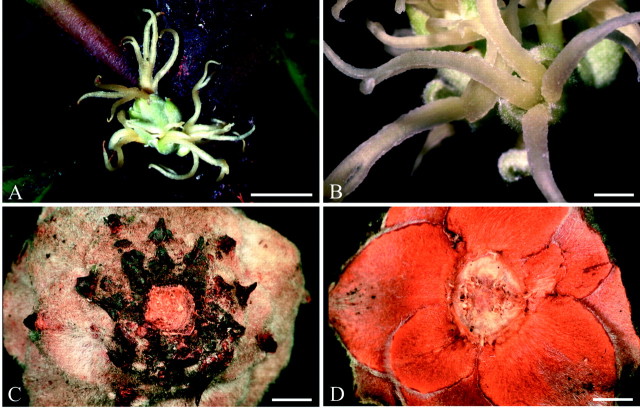
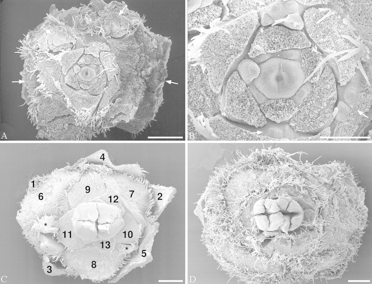
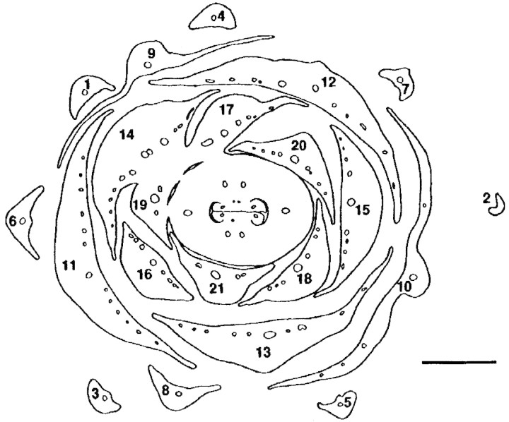
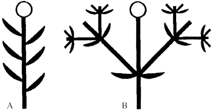
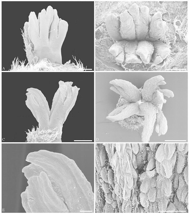
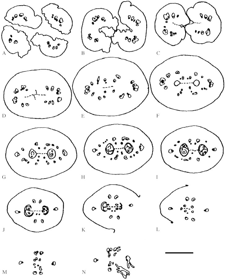
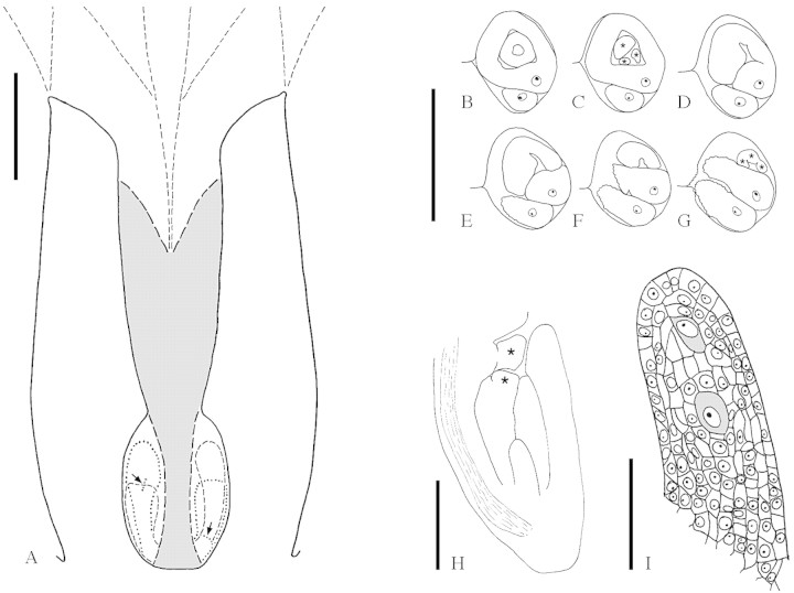
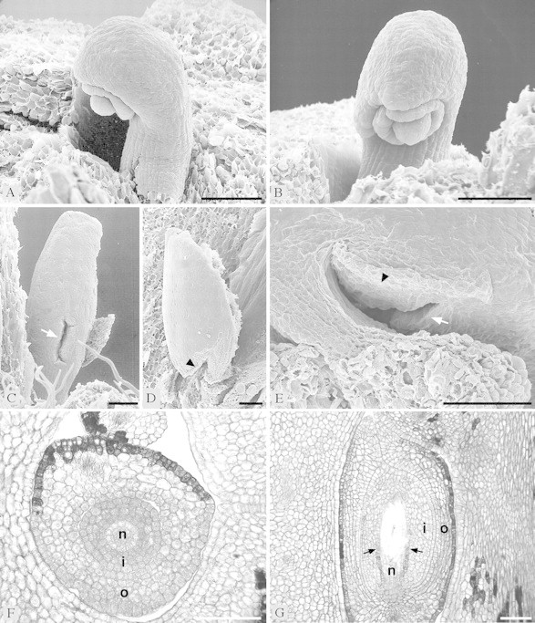
The Balanopaceae, whose flowers were poorly known, have, in the past, been variously allocated to the Fagales, Euphorbiaceae, Salicales or other hamamelids and rosids (these groups being in Fagales, Malpighiales and Saxifragales, according to the Angiosperm Phylogeny Group). This paper attempts a clarification based on flower morphology. Female flowers and cupules were studied in Balanops vieillardii, young fruits in B. australiana. The cupules are simple involucres of bracts which are spirally arranged (according to a Fibonacci pattern) on the floral axis preceding the flower. They contrast with the complicated cupules of Fagaceae which consist of a condensed cymose ramification system of axes of several orders around the flower. Flowers appear later than most of the cupular bracts, in contrast to Fagaceae. In addition to a terminal flower there may be several smaller lateral flowers in the axil of cupular bracts, each surrounded by its own small cupule. The female flowers do not have a perianth. They consist of two to three large carpels. At anthesis, the ovary is completely septate; the syncarpous part (ovary and lower style) is completely symplicate. The carpels are free for most of their length, with the free parts once, twice or three times bifurcate, in contrast to simple in Fagales. The stigmatic surface covers the ventral side of each stigmatic branch and at the margins also spreads to the dorsal side. The stigma is wet and secretion appears holocrine. The two ovules per carpel are collateral and axile in early development. However, at anthesis they appear one above the other, because in one ovule the funicle greatly elongates. As the ovary elongates only above the placenta, the ovules appear basal at anthesis. The ovules are (weakly) crassinucellar, bitegmic (not unitegmic), anatropous, and intermediate between apotropous and epitropous (not apotropous). The ovules are mature at anthesis, in contrast to Fagales. In mature ovules the upper part of the nucellus disintegrates, and a weakly differentiated endothelium is present in the inner integument. The morphological results of this study support a position of Balanopaceae in Malpighiales, and not Fagales or other orders, and are thus in accordance with recent molecular results based on chloroplast rbcL sequences data. However, within Malpighiales, as opposed to molecular results, Balanopaceae agree more with Euphorbiaceae s.l. than with Dichapetalaceae/Trigoniaceae and Chrysobalanaceae/Euphroniaceae.


Figures

None
Fig. 1.Balanopsvieillardii. Inflorescence and cupule of fruit: A, inflorescence, terminal flower above, lateral flowers below; B, lateral flower; C, fruit cupule from below; D, fruit cupule from above. Bars: A = 1 cm; B–D = 2 mm.
None
Fig. 2.Balanopsvieillardii. Young inflorescence and terminal flowers. Bracts removed to show flowers. A, Overview of inflorescence with terminal and lateral flowers. B, Terminal flower of the same inflorescence, at higher magnification. C, Terminal flower, older, showing first and second bifurcation of carpels. Subsequent bracts of cupule numbered to show spiral arrangement. D, Terminal flower, showing first, second and third bifurcation of carpels. White arrows: reduced lateral flowers; stars: lateral buds. Bars: A, C and D = 0·5 mm; B = 0·25 mm.
None
Fig. 3.Balanopsvieillardii. Transverse section of cupule with terminal female flower at the level at which the flower and the uppermost bracts of the cupule fuse. Subsequent bracts of cupule numbered to show spiral arrangement. Bar = 1 mm.
None
Fig. 4. Diagrams of cupules: A, Balanopaceae; B, Fagaceae. In the diagrams the axes are much longer than they are in reality to illustrate the ramification pattern. Flowers are represented by circles. Axes and bracts that are constituents of the cupule are represented by thick black symbols.
None
Fig. 5.Balanops vieillardii. Female flowers at different developmental stages. A and B, Younger preanthetic flower: A, from the side; B, from above. C–E, More advanced preanthetic flower: C, from the side; D, from above; E, close-up of C, showing dorsal side of stigmatic lobes with bulging stigmatic tissue at the margins. F, Part of stigma of anthetic flower, epidermal cells secretory and collapsing. Bars: A and B = 0·5 mm; C and D = 1 mm; E = 0·2 mm; F = 0·1 mm.
None
Fig. 6.Balanops vieillardii. TS series of somewhat preanthetic flower. A–C, Apocarpous zone: A, level of first bifurcation of free carpels; B, below first bifurcation, ventral slits open; C, ventral slits closed. D–L, Symplicate zone: D and E, style; F–L, ovary, F, top of locule without ovules; G, upper level of longer ovules; H, micropylar level of longer ovules; I, micropylar level of one of the shorter ovules; J, funicular level of all ovules; K, placenta; L, lowermost section with one locule. M and N, Level below locules. Post-genitally fused areas are marked with interrupted lines. Vascular bundles (xylem black) marked with thinner lines. Bar = 1 mm.
None
Fig. 7.Balanops vieillardii. A, Schematic median LS of gynoecium at anthesis (upper stigmatic region not drawn). Post-genitally fused area is shaded. Outline of parts that are not exactly in the median plane interrupted. Micropyles are marked with arrows (longitudinal in longer ovule, transverse in shorter ovule). B–G, TS series of the two ovules in a locule, shorter ovule showing micropylar region, longer ovule showing funicle. Vascular bundle in raphe indicated (xylem black). Lobes of inner (in C) and outer (in G) integument marked with stars. B, Level of both integuments; C, lobes of inner integument; D–G, lobes of outer integument. H, Median LS ovule at megaspore mother cell stage. Vascular bundle in raphe indicated. Lobes of inner and outer integument marked with stars. I, Nucellus of the same ovule, showing two potential megaspore mother cells (shaded). Bars: A = 1 mm; B–G = 0·5 mm; H = 0·2 mm; I = 0·05 mm.
None
Fig. 8.Balanops vieillardii. Ovules. A and B, From preanthetic flower, showing two integuments and nucellus: A, from the side; B, from the front. C–E, From anthetic flower. Micropyle marked with white arrow. Edge around micropylar region where ovule is appressed to funicle marked with black arrowhead. C, Longer ovule from the front, showing micropyle; D–E, shorter ovule; D, from the side, with microyple appressed to funicle; E, obliquely from below, showing micropyle. F–G, Microtome sections of ovules (n, nucellus; i, inner integument; o, outer integument): F, TS of preanthetic ovule; G, LS of anthetic ovule, with nucellus almost disintegrated by growing embryo sac. Weakly differentiated endothelium marked with arrows. Bar = 0·1 mm.

Similar articles

Cited by

References

    1. APG(The Angiosperm Phylogeny Group).1998. An ordinal classification for the families of flowering plants. Annals of the Missouri Botanical Garden 85: 531–553.
    1. BaillonH.1871. Sur deux nouveaux genres apétales. Adansonia 10: 112–119.
    1. BaillonH.1872. Stirpes exoticae novae (continué). Adansonia 10: 334–345.
    1. BaillonH.1877.Histoire des plantes VI. Paris: Hachette.
    1. BenthamG.1880. Balanopseae. In: Bentham G, Hooker JD, eds. Genera plantarum, Vol. 3. London: Reeve, 341.

MeSH terms