. 2003 Aug 20;23(20):7543-50.
doi: 10.1523/JNEUROSCI.23-20-07543.2003.

Circadian clock-controlled regulation of cGMP-protein kinase G in the nocturnal domain

Affiliations

Circadian clock-controlled regulation of cGMP-protein kinase G in the nocturnal domain

Shelley A Tischkau et al. J Neurosci. .

Abstract

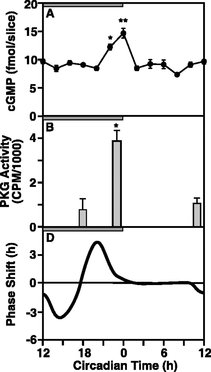
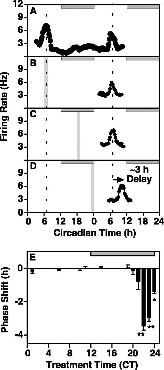
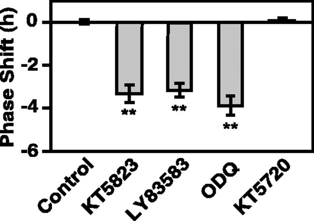
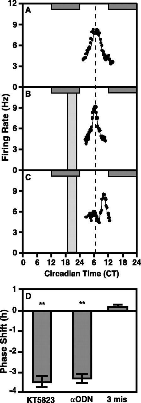
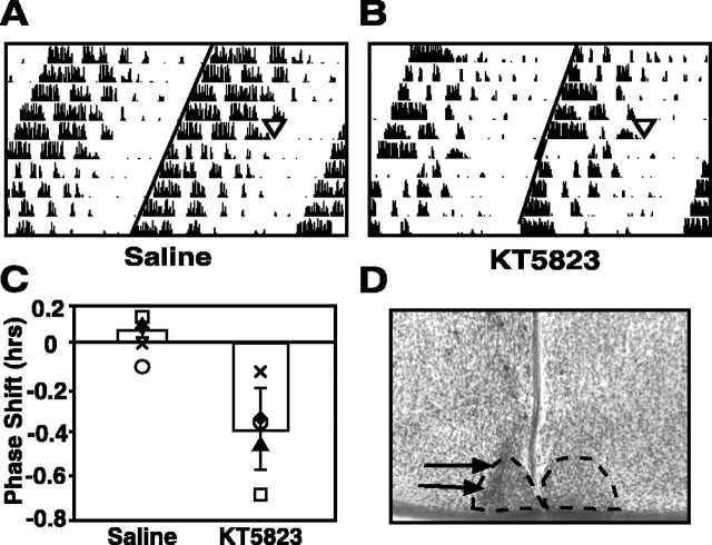
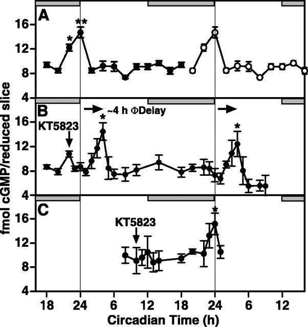
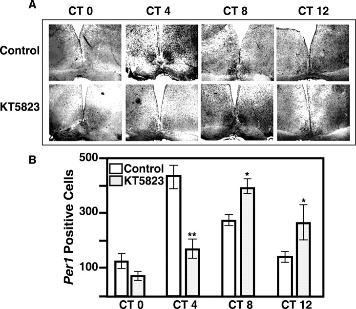
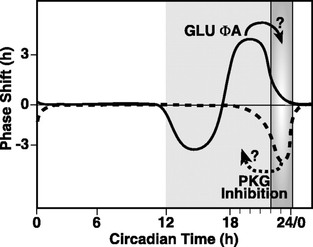
The suprachiasmatic nucleus (SCN) circadian clock exhibits a recurrent series of dynamic cellular states, characterized by the ability of exogenous signals to activate defined kinases that alter clock time. To explore potential relationships between kinase activation by exogenous signals and endogenous control mechanisms, we examined clock-controlled protein kinase G (PKG) regulation in the mammalian SCN. Signaling via the cGMP-PKG pathway is required for light- or glutamate (GLU)-induced phase advance in late night. Spontaneous cGMP-PKG activation occurred at the end of subjective night in free-running SCN in vitro. Phasing of the SCN rhythm in vitro was delayed by approximately 3 hr after treatment with guanylyl cyclase (GC) inhibitors, PKG inhibition, or antisense oligodeoxynucleotide (alphaODN) specific for PKG, but not PKA inhibitor or mismatched ODN. This sensitivity to GC-PKG inhibition was limited to the same 2 hr time window demarcated by clock-controlled activation of cGMP-PKG. Inhibition of the cGMP-PKG pathway at this time caused delays in the phasing of four endogenous rhythms: wheel-running activity, neuronal activity, cGMP, and Per1. Timing of the cGMP-PKG-necessary window in both rat and mouse depended on clock phase, established by the antecedent light/dark cycle rather than solar time. Because behavioral, neurophysiological, biochemical, and molecular rhythms showed the same temporal sensitivities and qualitative responses, we predict that clock-regulated GC-cGMP-PKG activation may provide a necessary cue as to clock state at the end of the nocturnal domain. Because sensitivity to phase advance by light-GLU-activated GC-cGMP-PKG occurs in juxtaposition, these signals may induce a premature shift to this PKG-necessary clock state.


Figures

Figure 1.
Figure 1.
Relationships between oscillations in cGMP levels and PKG activity and the phase response curve for GLU, which relates the time of GLU stimulation to the resultant effect on clock phase observed in rat SCN maintained in vitro. The horizontal gray bar indicates nighttime in the donor colony; slices were maintained in constant light. A, cGMP levels undergo a significant oscillation. Whereas cGMP levels were low during subjective day and most of subjective night, cGMP was significantly higher (ANOVA) at CT 22 (p < 0.05) and CT 24 (p < 0.01) compared with all other times. Data are presented as mean ± SEM for seven experiments. Two rat SCN were pooled within each experiment for every time point. B, In vitro phosphorylation of the specific PKG substrate RKRSRAE reflects the endogenous PKG activity of rat SCN obtained at CT 18, 23, and 11. PKG activity in samples containing 1.0 μm KT5823 was subtracted from activity in uninhibited samples to yield specific PKG activity. PKG specific activity (CPM per reduced slice) was significantly higher at CT 23 compared with CT 11 and 18 (ANOVA; p < 0.05). Data are means ± SEM for four experiments. C, The phase response curve for the effect of GLU on the timing of the oscillation in rat SCN neuronal activity, showing maximum sensitivity to phase advance at CT 19-20 (redrawn from Ding et al., 1994). *Statistical significance, as determined by ANOVA and the Student—Neuman-Keuls post hoc test, indicated by *p < 0.05, **p < 0.01, throughout, except Figure 5.
Figure 2.
Figure 2.
Inhibition of PKG causes a significant phase delay in the electrical activity rhythm of the rat SCN in vitro. The horizontal gray bar indicates nighttime in the donor colony; slices were maintained in constant light. Vertical bars indicate the time and duration of treatment. A, A representative single-unit recording from the ensemble of SCN neurons shows an endogenous electrical activity rhythm with a peak at ∼CT 7 on both day one and day two in vitro. Mean time-of-peak for control experiments (n = 8) was CT 6.88 ± 0.11. B, A 1 hr bath application of the specific PKG inhibitor, KT5823 (1.0 μm), at CT 6 had no effect on the time-of-peak of the SCN electrical activity rhythm on the subsequent day. Data shown are from a representative experiment. The average time-of-peak was CT 6.99 ± 0.23 (n = 3), which is not significantly different from control (Student's t test). C, A 1 hr bath application of KT5823 (1.0 μm) at CT 18 had no effect on the time-of-peak of the SCN electrical activity rhythm on the subsequent day. Data shown are from a representative experiment. The average time-of-peak was CT 6.90 ± 0.45 (n = 3), which was not significantly different from control (Student's t test). D, A 1 hr bath application of KT5823 (1.0 μm) at CT 23 induced a significant phase delay in the SCN electrical activity rhythm (p < 0.01; Student's t test). Data shown are from a representative experiment. The mean time-of-peak was delayed by 3 hr, shifting from CT 6.88 ± 0.11 (n = 7) to CT 9.83 ± 0.24 (n = 4). E, The effects of PKG inhibition are temporally restricted to a narrow window of sensitivity during the late night/predawn period. KT5823 was applied to the bath for 1 hr at 11 different points on the circadian cycle (CT 2, CT 6, CT 10, CT 11, CT 14, and CT 18-23). Each bar represents the mean ± SEM of 3-6 experiments. Sensitivity to phase delay induced by KT5823 was restricted to the end of subjective night. Significant phase delays were observed when the inhibitor was applied at CT 22 (-3.46 ± 0.24; n = 6; p < 0.01), CT 23 (-2.96 ± 0.24; n = 4; p < 0.01), and CT 24 (-1.38 ± 0.14; n = 3; p < 0.05).
Figure 3.
Figure 3.
Sensitivity to phase delay at the end of subjective night is selective for inhibition of the GC-cGMP-PKG-dependent pathway. Similar to the effects of KT5823 (replotted from Fig. 2), the GC inhibitors LY83583 and ODQ caused phase delays of rat SCN electrical activity rhythms when applied at CT 22. LY83583 (2 μm) induced a 3.2 ± 0.32 hr (n = 3; p < 0.01) delay, whereas the delay induced by ODQ (20 nm) was 3.88 ± 0.45 hr (n = 3; p < 0.01). The PKA inhibitor KT5720 (100 nm) had no effect on the electrical activity rhythm when applied at CT 23 (mean time-of-peak, CT 6.68 ± 0.33; n = 3). Duration of each treatment was 1 hr.
Figure 4.
Figure 4.
Inhibition of PKG using αODN causes a significant phase delay in the electrical activity rhythm of the rat SCN in vitro. The horizontal gray bar indicates nighttime in the donor colony; slices were maintained in constant light. Vertical bars indicate the time and duration of treatment. A, A representative single unit recording from the ensemble of SCN neurons shows an endogenous electrical activity rhythm with a peak at approximately CT 7 on both days one and two in vitro. Mean time-of-peak for control experiments (n = 8) was CT 6.88 ± 0.11. B, A 4 hr bath application of the PKG αODN (10 μm) bearing three mismatched nucleotides from CT 18-23 had no effect on the time-of-peak of the SCN electrical activity rhythm on the subsequent 2 d. Data shown are from a representative experiment. The average time-of-peak was CT 6.75 ± 0.25 (n = 3), which is not significantly different from control (Student's t test). C, A 4 hr bath application of PKG αODN (10 μm) from CT 19-23 induced a significant phase delay in the SCN electrical activity rhythm (p < 0.01; Student's t test). Data shown are from a representative experiment. The mean time-of-peak was delayed by 3.12 hr, shifting from CT 6.88 ± 0.11 (n = 7) to CT 9.00 ± 0.54 (n = 4). D, Summary of PKG ODN effects. αODN treatments caused phase delays equal in magnitude to those observed after treatment with KT5823, whereas a three base mismatch ODN did not alter phase (replotted from Fig. 2).
Figure 5.
Figure 5.
Inhibition of PKG at the end of subjective night induces phase delays in vivo in the mouse. A, B, Representative double-plotted actograms depicting effects of intra-SCN injections at CT 0 of saline (A) (0.3 μl, 0.9% NaCl) or KT5823 (B) (0.3 μl, 100 μm). Each horizontal line indicates 48 hr of data, with the last 24 hr of each line replotted as the first 24 hr of the following line. Treatments are indicated with triangles, and vertical black lines indicate the relative magnitude of running wheel activity. Diagonal lines have been drawn to aid in visualization of the phase shifts. C, A bar graph depicting average phase shifts ± SEM after indicated treatments. Shapes represent individual subject's responses and are paired across treatment conditions. Phase shifts were determined by calculating the difference in hours between two regression lines; one plotted through the 5 d of activity onsets preceding treatment, and the other plotted through the 5 d of activity after a return to stable running-wheel patterns after treatment. There were five animals in each treatment condition. Responses were determined to be significantly different (p < 0.01) by Student's paired t test. D, Representative histology of the intra-SCN injection site. The SCN have been outlined with a dotted line, and arrows point to the location the cannula.
Figure 6.
Figure 6.
Inhibition of PKG at the end of subjective night induces phase delays in the rat cGMP oscillation. A, cGMP levels in SCN slices maintained in vitro oscillate with a peak at CT 24. These data are double-plotted for reference of the basal rhythms over 2 d (data from Fig. 1). *p < 0.05 and **p < 0.01 indicate samples that are statistically different compared with CT 20 values. Significance was determined by ANOVA and Student-Neuman-Keuls test. B, A 15 min bath application of KT5823 at CT 22 causes cGMP levels to return to basal (CT 20) levels within 1 hr and causes a significant (p < 0.01; Student's t test) phase delay in the cGMP oscillation. The shift in cGMP persists for 2 d in vitro. Data are means ± SEM for four separate experiments. C, KT5823 had no effect on the cGMP oscillation when applied for 15 min to the bath at CT 10. Individual data points are means ± SEM for four separate experiments.
Figure 7.
Figure 7.
Inhibition of PKG at the end of subjective night alters the expression pattern of Per1 mRNA in the rat SCN. A, Representative in situ hybridization histochemistry. In control slices, a significant increase in Per1 mRNA was observed with a peak at CT 4. After KT5823 treatment for 1 hr at CT 22, the endogenous rise in Per1 was delayed by 4 hr; PKG-treated slices showed peak Per1 expression at CT 8. Magnification, 200×. B, Quantitation of A. Per1-positive cells were counted by an experimenter blind to the experimental treatments. Data represent the mean ± SEM of four independent experiments. Statistical analysis (ANOVA with Student-Neuman-Keuls post hoc analysis; p < 0.01) revealed a significant increase in Per1 mRNA at CT 4 and CT 8 in control slices, with a peak at CT 4. In KT5823-treated slices, a significant increase was observed at CT 8 and CT 12 with a peak at CT 8. CT 4 controls had significantly more Per1-positive cells than CT 4-treated slices (p < 0.01; Student's t test). Treated slices had significantly more Per1-positive cells at CT 8 (p < 0.05) and CT 12 (p < 0.01), compared with time-matched controls.
Figure 8.
Figure 8.
Model of circadian clock regulation by cGMP-PKG at the end of subjective night. The SCN is sensitive to phase resetting by stimuli dependent on cGMP and PKG activation during the last half of subjective night. These stimuli cause phase advances that may be manifested as shifting clock state forward several hours to the end of the night (solid arrow). The domain of endogenous rise in cGMP and PKG activity (gradient gray) is a narrow window of time that is coincident with the waning of sensitivity to phase advance stimulated by light-GLU via cGMP-PKG-dependent mechanisms. Inhibiting PKG activity during this time causes a 3 hr phase delay. This may be a dynamic process that shifts the clock back in time, into the domain of sensitivity to phase resetting by exogenous stimuli that activate the cGMP-PKG pathway (dashed arrow). The tight temporal relationship between exogenous sensitivity and endogenous activation of cGMP-PKG suggests that light-GLU may prematurely stimulate a system poised to respond ∼3.5 hr later to clock-controlled processes. The clock-controlled rise in cGMP levels and concomitant activation of PKG at the end of the night comprise a critical event in this time domain.

Similar articles

Cited by

References

    1. Albrecht U, Sun ZS, Eichele G, Lee CC ( 1997) A differential response of two putative mammalian circadian regulators, mper1 and mper2, to light. Cell 91: 1055-1064. - PubMed
    1. Balsalobre A, Damiola F, Schibler U ( 1998) A serum shock induces circadian gene expression in mammalian tissue culture cells. Cell 93: 929-937. - PubMed
    1. Best JD, Maywood ES, Smith KL, Hastings MH ( 1999) Rapid resetting of the mammalian circadian clock. J Neurosci 19: 828-835. - PMC - PubMed
    1. Comolli JC, Hastings JW ( 1999) Novel effects on the Gonyaulax circadian system produced by the protein kinase inhibitor staurosporine. J Biol Rhythms 14: 11-19. - PubMed
    1. Crosthwaite SK, Dunlap JC, Loros JJ ( 1997) Neurospora wc-1 and wc-2: transcription, photoresponses, and the origins of circadian rhythmicity. Science 276: 763-769. - PubMed

Publication types

MeSH terms

LinkOut - more resources