. 2003 Aug 20;23(20):7602-9.
doi: 10.1523/JNEUROSCI.23-20-07602.2003.

Semaphorin 3F antagonizes neurotrophin-induced phosphatidylinositol 3-kinase and mitogen-activated protein kinase kinase signaling: a mechanism for growth cone collapse

Affiliations

Semaphorin 3F antagonizes neurotrophin-induced phosphatidylinositol 3-kinase and mitogen-activated protein kinase kinase signaling: a mechanism for growth cone collapse

Jasvinder K Atwal et al. J Neurosci. .

Abstract

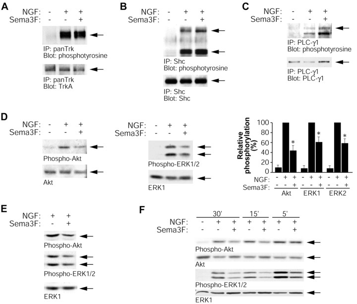
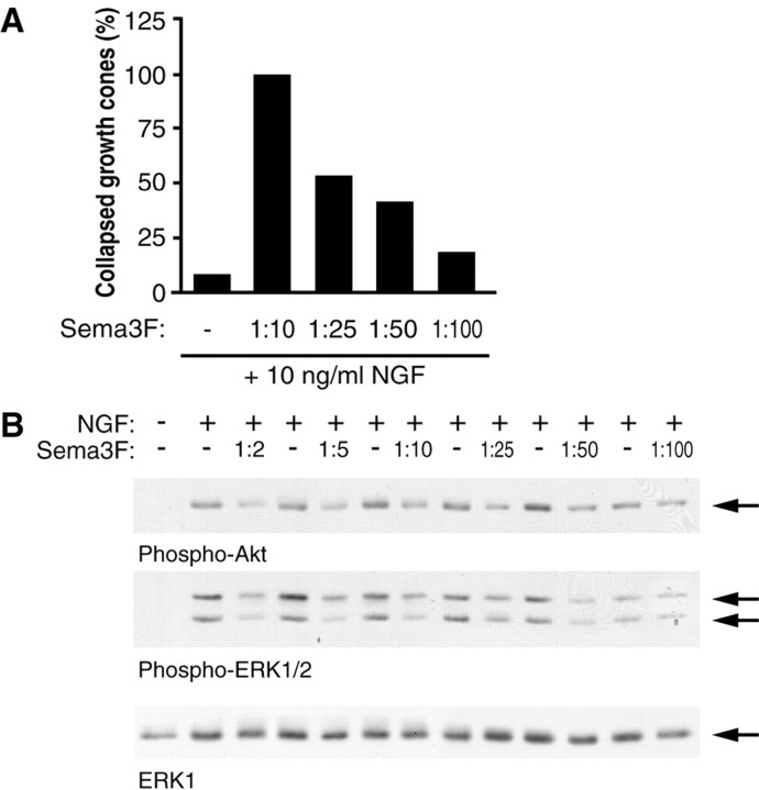
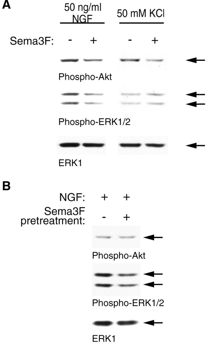
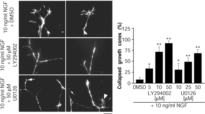
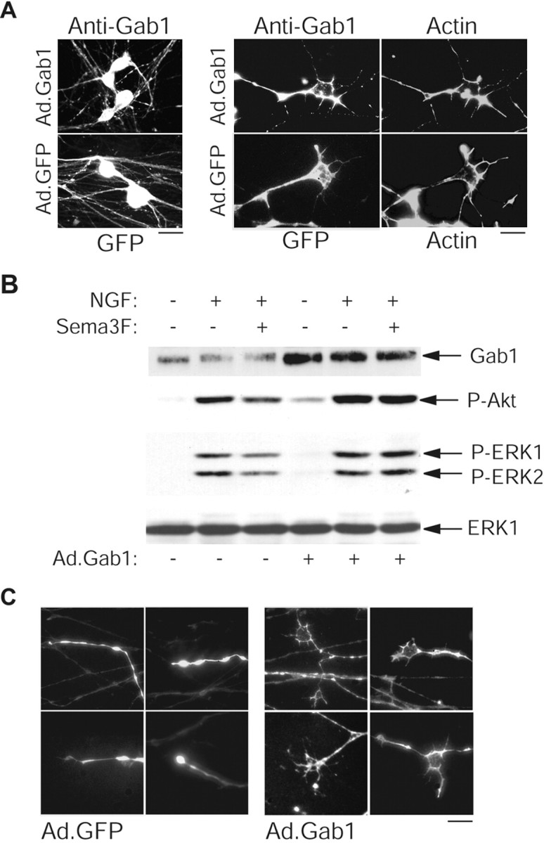
Peripheral nerve growth is regulated by the coordinated action of numerous external stimuli, including positively acting neurotrophin-derived growth cues and restrictive semaphorin cues. Here, we show that Semaphorin 3F (Sema 3F) can antagonize nerve growth factor (NGF)-stimulated TrkA (tyrosine receptor kinase A) signaling in sympathetic neurons, thereby apparently contributing to growth cone collapse. Sema 3F suppressed NGF-induced activation of the phosphatidylinositol 3 (PI3)-kinase-Akt and MEK (mitogen-activated protein kinase kinase)-ERK (extracellular signal-regulated kinase) pathways, both of which we show to be required to maintain growth cone structure. Sema 3F-induced growth cone collapse was partially reversed by sustained activation of the PI3-kinase and MEK pathways, which was achieved by overexpression of the Gab-1 (growth-associated binder 1) docking protein. These data indicate that a novel mechanism used by Sema 3F to collapse growth cones in sympathetic neurons is to dampen neurotrophin signaling, providing an intracellular mechanism for cross talk between positive and negative axon growth cues.


Figures

Figure 1.
Figure 1.
Postnatal sympathetic neurons express TrkA and Npn-2 in growth cones. A, Pan-Trk or Npn-2 antibodies were used to immunoprecipitate (IP) proteins from sympathetic neuron lysates. Western blotting (Blot) was then performed to show the presence of both TrkA (anti-RTA) and Npn-2. Arrows indicate bands of the appropriate size. B, TrkA and Npn-2 expression in growth cones was examined by comparing immunoreactive regions with actin-rich growth cones, as detected by rhodamine-phalloidin costaining. Both TrkA and Npn-2 expression are found within growth cones. Scale bar, 5 μm.
Figure 2.
Figure 2.
Growth cone collapse is caused by Sema 3F and inhibition of NGF-TrkA signaling. A, Schematic diagram of Campenot chambers, in which cells are plated in the center compartment, and axons grow into side compartments. In experiments described here, side compartments contained 10 ng/ml of NGF, axons were acutely treated for 30 min at 37°C under different conditions as indicated, and growth cones were analyzed by rhodamine-phalloidin staining. B, Sema 3F collapses NGF-induced growth cones. Left, Representative micrographs of rhodamine-phalloidin-stained growth cones treated with 10 ng/ml of NGF or 10 ng/ml of NGF plus Sema 3F. The graph indicates the fraction of collapsed growth cones for the two conditions (n = 4). C, Sema 3F inhibits long-term axonal growth. Left, Representative micrographs showing axonal density in compartments treated for 2 d with 10 ng/ml of NGF plus or minus Sema 3F. Right, A Western blot for total α-tubulin in compartments treated under these two conditions. The arrow indicates a band of the appropriate size. D, Inhibition of NGF or TrkA causes growth cone collapse. Photographs are representative micrographs of rhodamine-phalloidin-stained growth cones treated with anti-NGF (1:1000), K252a (200 nm), or DMSO as a control. The graph indicates the fraction of collapsed growth cones for each condition (n = 3). Scale bars: B, D, 10 μm; C, 80 μm. *p < 0.001; t test comparing treatment to NGF (B) or NGF plus DMSO (D).
Figure 3.
Figure 3.
Sema 3F-mediated collapse of sympathetic growth cones is not antagonized by inhibition of Rho-kinase or elevation of cGMP. A, B, Axonal side compartments from compartmented cultures of sympathetic neurons were treated for 30 min with 10 ng/ml of NGF, 10 ng/ml of NGF plus Sema 3F, or 10 ng/ml of NGF plus Sema 3F with either 10 μm Y26732 (A) or 5 mm 8-Br-cGMP (B). Photographs are representative micrographs of rhodamine-phalloidin-stained growth cones after the various treatments, showing that neither Y27632 nor 8-Br-cGMP could inhibit the Sema 3F-mediated collapse. The graphs indicate the fraction of collapsed growth cones for each condition (n = 3 for Y27632; n = 2 for 8-Br-cGMP). Scale bars, 10 μm.
Figure 4.
Figure 4.
Sema 3F treatment affects downstream TrkA-signaling pathways. Mass cultures of sympathetic neurons were stimulated for 30 min with medium alone, 50 ng/ml of NGF, or 50 ng/ml of NGF plus Sema 3F, and cells were lysed and analyzed for activation of proteins in the Trk-signaling pathway. A-C, Tyrosine phosphorylation of TrkA (A), Shc (B), and PLC-γ1 (C) was assessed by immunoprecipitating with antibodies specific to each protein and probing Western blots with anti-phosphotyrosine. Arrows indicate TrkA (A), multiple Shc isoforms (B), or PLC-γ1 (C). Lower blots in each panel are reprobes of immunoprecipitated proteins. D, Total cell lysates were analyzed for Akt or ERK phosphorylation by Western blot, probing with phospho-Akt or phospho-ERK antibodies, and then reprobing for total Akt or ERK1. The graph shows the amount of Akt or ERK phosphorylation relative to the maximal phosphorylation seen with 50 ng/ml of NGF in the same experiment, averaged over five independent experiments. *p < 0.001; t test comparing NGF plus Sema 3F to NGF alone. E, Western blot analysis for phospho-Akt, phospho-ERK, or ERK1 reprobe in lysates from axons only in side compartments treated with 50 ng/ml of NGF or 50 ng/ml of NGF plus Sema 3F for 30 min. F, Time course of Sema 3F downregulation of Akt and ERK phosphorylation. Cells were washed and stimulated as described above and lysed after 5, 15, or 30 min of stimulation. Western blot analysis for phospho-Akt and phospho-ERK shows downregulation of ERK phosphorylation as early as 5 min after stimulation and downregulation of Akt phosphorylation by 15 min.
Figure 5.
Figure 5.
Dose-response curve of Sema 3F-mediated growth cone collapse and inhibition of Akt and ERK phosphorylation. A, Axons in compartmented cultures were treated with 10 ng/ml of NGF plus or minus varying dilutions of Sema 3F-conditioned medium and the percentage of collapsed growth cones quantitated by rhodamine-phalloidin staining after 30 min. B, Mass cultures of sympathetic neurons were treated with 50 ng/ml of NGF plus or minus varying dilutions of Sema 3F-conditioned medium, and Western blot analysis was used to quantitate the extent of Akt and ERK phosphorylation using phosphorylation-specific antibodies. The blots were then reprobed for total ERK1 to evaluate protein levels. Similar results were obtained in two independent experiments. Arrows indicate bands of appropriate sizes.
Figure 6.
Figure 6.
Sema 3F can inhibit downstream Akt and ERK phosphorylation in a TrkA-independent manner. A, Mass cultures of sympathetic neurons were established in NGF and then switched to 50 mm KCl plus or minus 1:10 Sema 3F for 30 min. Alternatively, mass cultures of neurons were washed free of NGF and then switched to 1:10 Sema 3F for 25 min, washed again, and stimulated with 50 ng/ml of NGF alone for 5 min. Lysates were then analyzed by Western blot analysis for phospho-Akt, phospho-ERK, or total ERK1 protein. Similar results were obtained in two independent experiments. Arrows indicate bands of appropriate sizes.
Figure 7.
Figure 7.
Inhibition of the PI3-kinase-Akt or MEK-ERK pathways is sufficient to induce growth cone collapse in sympathetic axons. Axons in side compartments were treated for 30 min with 10 ng/ml of NGF plus or minus LY294002 (5, 10, or 50 μm) to inhibit PI3-kinase, U0126 (10, 25, or 50 μm) to inhibit MEK, or DMSO as a control. Representative photomicrographs of rhodamine-phalloidin-stained growth cones are shown on the left. Quantification of growth cone collapse by the inhibitors is shown in the graph on the right (n = 4). *p < 0.05; **p < 0.001; t test comparing treatments to NGF plus DMSO.
Figure 8.
Figure 8.
Sustained activation of the PI3-kinase-Akt and MEK-ERK pathways by overexpressing Gab-1 partially rescues Sema 3F-induced growth cone collapse. Neurons were infected with 100 MOI recombinant adenovirus expressing wild-type Gab-1 (Ad.Gab-1) or GFP (Ad.GFP) overnight and analyzed 2 d after infection. A, Photomicrographs showing that GTP expression and Gab-1 immunoreactivity were detected in both cell bodies and axons (left) and could be visualized in growth cones, as seen by costaining with rhodamine-phalloidin-labeled actin (right). Endogenous Gab-1 was below the level of detectability by immunostaining. B, Overexpression of Gab-1 enhances PI3-kinase and ERK activation. Infected neurons were stimulated as described in Figure 3, and lysates were analyzed for Gab-1, phospho-Akt, phospho-Erk, or ERK1 levels. Arrows indicate bands of appropriate sizes. C, Representative photomicrographs of growth cones from Gab-1-expressing neurons (visualized by Gab-1 immunostaining) or GFP-expressing neurons (visualized directly) that were treated with 10 ng/ml of NGF plus Sema 3F for 30 min at 48 hr after infection. Scale bars: A, 15 μm (left), 5 μm (right); C, 10 μm.

References

    1. Aizawa H, Wakatsuki S, Ishii A, Moriyama K, Sasaki Y, Ohashi K, Sekine-Aizawa Y, Sehara-Fujisawa A, Mizuno K, Goshima Y, Yahara I ( 2001) Phosphorylation of cofilin by LIM-kinase is necessary for semaphorin 3A-induced growth cone collapse. Nat Neurosci 4: 367-373. - PubMed
    1. Arimura N, Inagaki N, Chihara K, Menager C, Nakamura N, Amano M, Iwamatsu A, Goshimia Y, Kaibuchi K ( 2000) Phosphorylation of collapsin response mediator protein-2 by Rho-kinase. Evidence for two separate signaling pathways for growth cone collapse. J Biol Chem 275: 23973-27980. - PubMed
    1. Atwal JK, Massie B, Miller FD, Kaplan DR ( 2000) The TrkB-Shc site signals neuronal survival and local axon growth via MEK and P13-kinase. Neuron 27: 265-277. - PubMed
    1. Behar O, Golden JA, Mashimo H, Schoen FJ, Fishman MC ( 1996) Semaphorin III is needed for normal patterning and growth of nerves, bones and heart. Nature 383: 525-528. - PubMed
    1. Cai H, Reed RR ( 1999) Cloning and characterization of neuropilin-1-interacting protein: a PSD-95/Dlg/ZO-1 domain-containing protein that interacts with the cytoplasmic domain of neuropilin-1. J Neurosci 19: 6519-6527. - PMC - PubMed

Publication types

MeSH terms

LinkOut - more resources