. 2003 Aug 20;23(20):7610-20.
doi: 10.1523/JNEUROSCI.23-20-07610.2003.

The meninges is a source of retinoic acid for the late-developing hindbrain

Affiliations

The meninges is a source of retinoic acid for the late-developing hindbrain

Jinghua Zhang et al. J Neurosci. .

Abstract

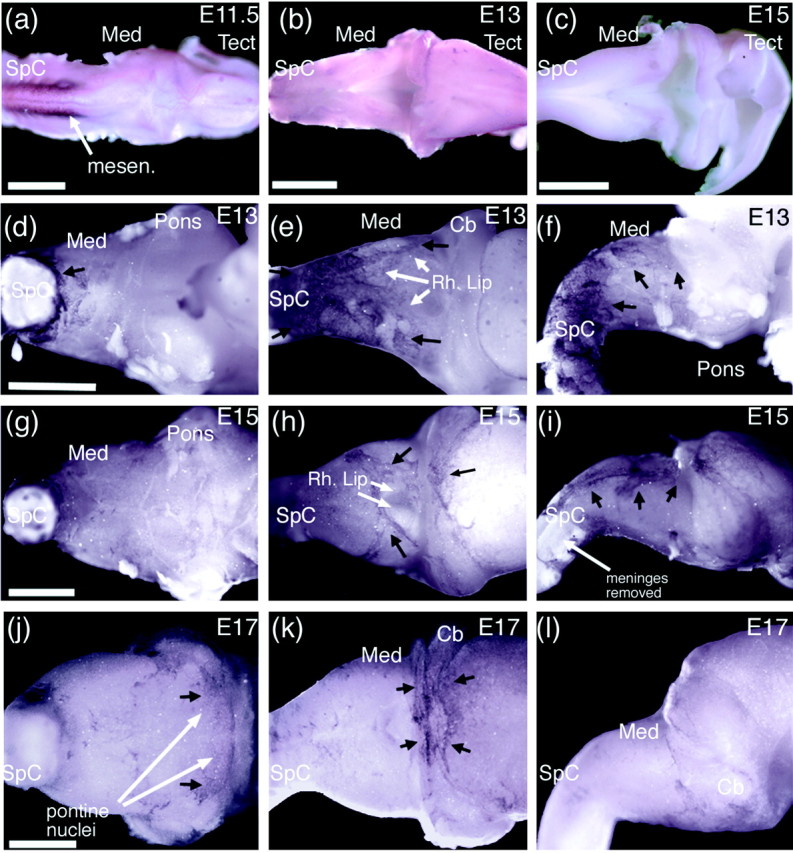
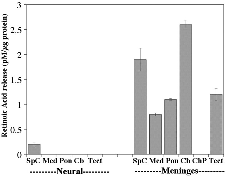
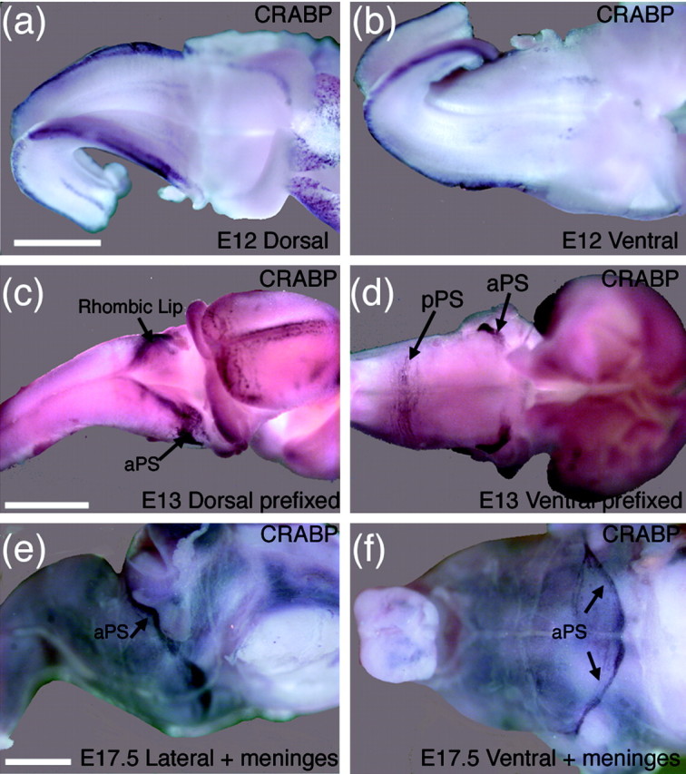
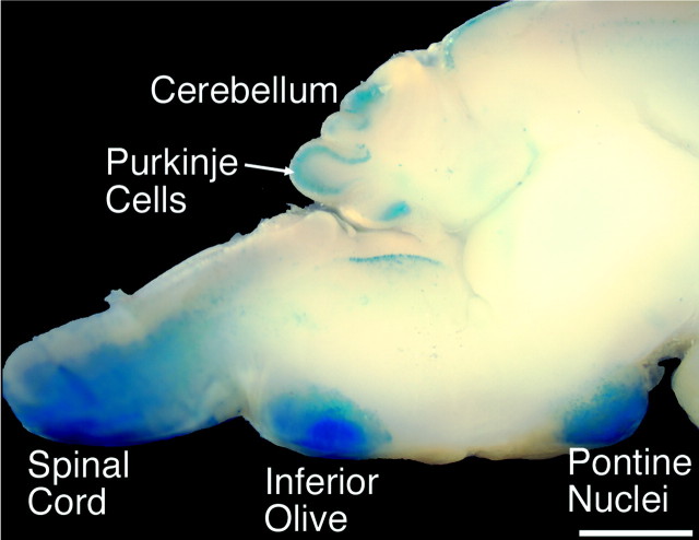
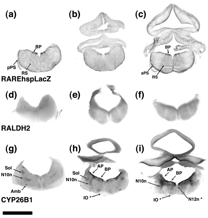
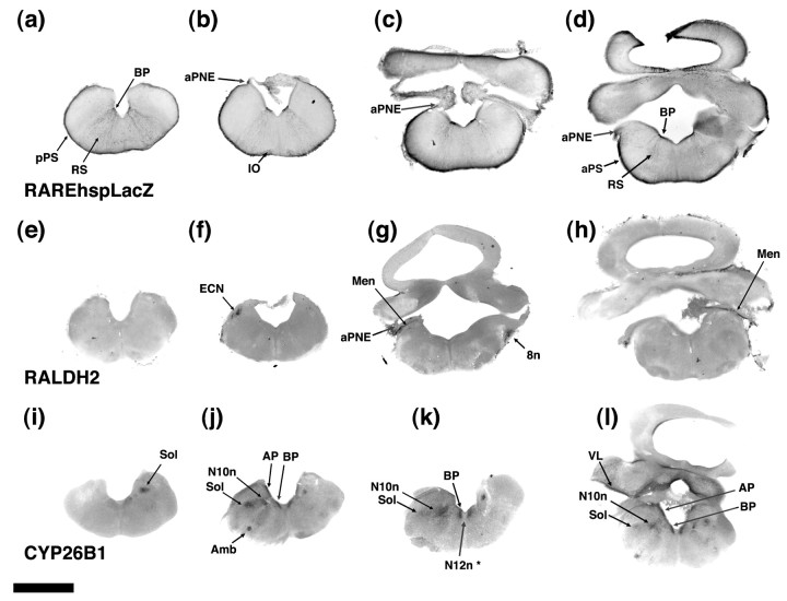
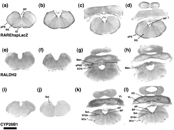
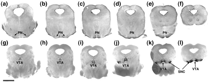
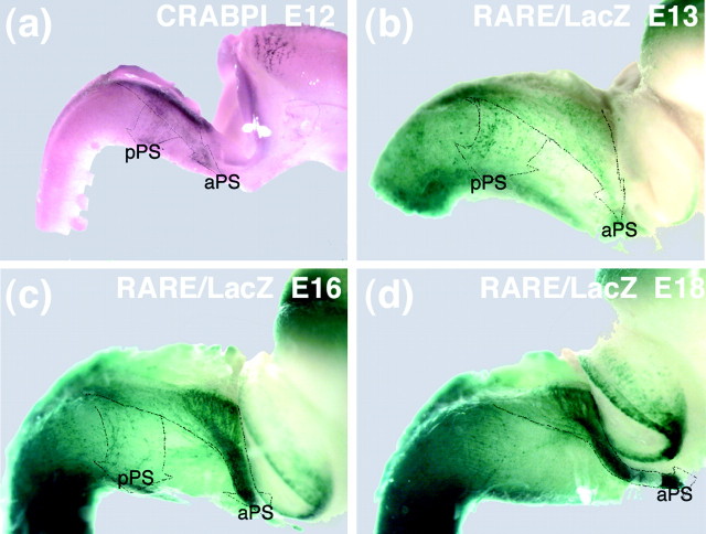
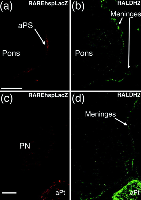
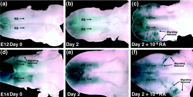
One general function for retinoic acid (RA) is pattern organization in the CNS. This regulatory factor has an essential role in spinal cord motor neuron and early posterior hindbrain development. In the anterior CNS, however, there is only a limited number of foci of RA synthesis, and less attention has been placed on regions such as the anterior hindbrain where RA synthesizing enzymes are absent. This study shows that a rich source of RA lies around the hindbrain from the RA synthetic enzyme retinaldehyde dehydrogenase-2 (RALDH2) present in the surrounding meninges and mesenchyme by embryonic day 13. RALDH2 is not distributed uniformly throughout the meninges but is restricted to territories over the developing hindbrain, suggesting that RA signaling may be localized to those regions. Further regulation of RA signaling is provided by the presence of a RA sink in the form of the CYP26B1 RA catabolic enzyme expressed in deeper regions of the brain. As a guide to the neural anatomy of hindbrain RA signaling, we used a mouse transgenic for a lacZ reporter gene driven by a RA response element (RAREhsplacZ) to identify regions of RA signaling. This reporter mouse provides evidence that RA signaling in the hindbrain after embryonic day 13 occurs in the regions of the cerebellum and precerebellar system adjacent to sources of RA, including the inferior olive and the pontine nuclei.


Figures

Figure 1.
Figure 1.
Pathways of migration of neurons born from the neuroepithelium of the rhombic lip.
Figure 2.
Figure 2.
In situ hybridization for RALDH2 in the developing hindbrain at E11.5 (a), E13 (b, d-f), E15 (c, g-i), and E17 (j-l) in dorsal (a-c, e, h, k), ventral (d, g, j), and lateral (f,i,l) views. At E11.5, RALDH2 is expressed in the mesenchyme (mesen.) surrounding the somites of the trunk (a) but is absent from the mesenchymal tissues over the dorsal brain. If the mesenchymal tissues and meninges are stripped from the brain, then very little RALDH2 is evident over the brain at E13 (b) or E15 (c). If the meninges is left intact, however, at E13, RALDH2 is clearly evident over the dorsal (e, f, black arrows) versus the ventral (d) surface and surrounds the dorsal rhombic lip. RALDH2 levels have fallen by E15 but are still higher in the meningeal network around the region of the rhombic lip (h, black arrows). Overall levels in the meninges are above those in the underlying CNS, indicated by the staining intensity in the spinal cord (i) where the meninges have peeled off (white arrow). At E 17, RALDH2 levels have risen in the region of the developing pontine nuclei (j, black arrows), and although RALDH2 is low in the dorsal medulla and rhombic lip, it is higher over the cerebellum and tectum (k, black arrow). Med, Medulla; Cb, cerebellum. Black arrows point to the outer edge of meningeal RALDH2 spread. Scale bars, 1 mm.
Figure 3.
Figure 3.
Relative amounts of RA released by the meninges and CNS at E16. Similar amounts of tissues were dissected from the spinal cord (SpC), pons (Pon), tectum (Tect), medulla (Med), and cerebellum (Cb). The meninges around these tissues was also dissected as well as the choroid plexus (ChPl) of the fourth ventricle. The tissues were incubated overnight in tissue culture medium, and the medium was assayed for content of RA using an RA reporter cell line. Absolute values were determined by comparison to an RA standard curve and plotted as a ratio of the total protein in the tissue. RA release from the spinal cord was detectable, whereas the hindbrain was entirely negative. The meninges released significant amounts of RA into the medium, the highest levels from the meninges around the cerebellum.
Figure 4.
Figure 4.
Whole-mount in situ hybridization of CRABPI in hindbrain at E12 (a, b) and E13 (c, d) without meninges and at E17.5 with meninges (e,f). At E12 with the meninges removed before fixation (a) CRABPI is initially expressed along the dorsal edge of the hindbrain and the posterior edge of the cerebellum. If the brain is first fixed, then fewer of the neurons migrating under the meninges are lost when the meninges is removed and labeling of the rhombic lip is clearly evident (c) together with the stream of cells leaving the anterior precerebellar epithelium as part of the anterior precerebellar stream (aPS) moving toward (but not reaching at this age) the pontine nuclei. A minor stream of cells around the posterior medulla (d), the posterior precerebellar stream (pPS, arrow), is likely a second stream of cells derived from the posterior precerebellar neuroepithelium that migrate under the meninges to populate the external cuneate and lateral reticular nuclei. If the meninges are left on at E17.5, the anterior precerebellar stream (aPS) is clearly evident and has reached the pontine nuclei (e, f). Scale bars, 1 mm.
Figure 5.
Figure 5.
RAREhsplacZ expression in a sagittal section of postnatal day 3 hindbrain. A glutaraldehyde-fixed postnatal day 3 hindbrain was sliced immediately lateral to the midline and stained for β-galactosidase activity. In the hindbrain, the inferior olive and pontine nuclei are particularly strongly labeled as well as the Purkinje cells (PC) in the cerebellum. Scale bar, 1 mm.
Figure 6.
Figure 6.
Comparison of RAREhsplacZ, RALDH2, and CYP26B1 in the E13 hindbrain without meninges. RAREhsplacZ (a-c) expression is almost all distributed around the peripheries of the hindbrain in migrating cells of the posterior (pPS) and anterior (aPS) precerebellar stream and in the basal plate (BP), and the only labeled region within the brain is a stream of cell arising from the basal plate labeled the radial stream (RS). RALDH2 (d-f) is entirely absent from the brain, whereas CYP26B1 (g-i) is strongly expressed in the ventricular layer of the alar plate (AP) and basal plate (BP) and is also present in the region of the nucleus of the solitary tract (Sol), the dorsal nucleus of the vagus (N10n), and ambiguus nucleus (Amb). Several nuclei (labeled with asterisks) do not express CYB26B1, but the enzyme lies immediately adjacent to the cell group, and this includes the inferior olive (IO *) and nuclei of the hypoglossal nerve (N12n *). Scale bar, 1 mm.
Figure 7.
Figure 7.
Comparison of RAREhsplacZ, RALDH2, and CYP26B1 in the E15 hindbrain without meninges. As found at E13, RAREhsplacZ (a-d) continues to be induced in the peripheries of the hindbrain in the posterior (pPS) and anterior (aPS) precerebellar stream, basal plate (BP), and cells moving out from the basal plate in the radial stream (RS) but is absent from the anterior precerebellar neuroepithelium (aPNE). RALDH2 (e-h) is present in meninges (Men) that remained trapped between the aPNE and medulla but is also in two regions of the brain, the external cuneate nucleus (ECN) and vestibular nerve (8n). CYP6B1 (i-l) continues to be expressed in the ventricular layer of the alar plate (AP) and basal plate (BP) as well as the cerebellar ventricular layer (VL) and is also in the nucleus of the solitary tract (Sol), the dorsal motor nucleus of the vagus (N10n), and nucleus ambiguus (Amb) while the enzyme is expressed adjacent to the nuclei of the hypoglossal nerve (N12n *). Scale bar, 1 mm.
Figure 8.
Figure 8.
Comparison of RAREhsplacZ, RALDH2, and CYP26B1 in the E17 hindbrain without meninges. RAREhsplacZ (a-d) continues to be in the peripheries of the hindbrain in the posterior precerebellar stream (pPS), which merges into the ventral aspects of the inferior olive (IO) as well as the anterior precerebellar stream (aPS). The basal plate (BP) and radial stream (RS) also remain positive. RALDH2 (e-h) is present in meninges (Men) trapped between the precerebellar neuroepithelium (aPNE) and medulla and is also in the external cuneate nucleus (ECN). CYP26B1 (i-l) is in the ventricular layer (VL) of the cerebellum where it is also expressed in the Purkinje cell layer (PC). In the medulla CYP26B1 is in the ventricular layer of the alar plate (AP) and basal plate (BP), the solitary nucleus (Sol), the dorsal motor nucleus of the vagus (N10n), and adjacent to the nucleus of the facial nerve (N7n*). Scale bar, 1 mm.
Figure 10.
Figure 10.
Relative distribution of RAREhsplacZ and RALDH1 in the boundary between the pons and midbrain. Serial 100 μm vibratome sections labeled by immunohistochemistry for RAREhsplacZ (a-f) and by in situ hybridization for RALDH1 (g-l) aligning the approximate corresponding section for the two probes with the most caudal section on the left. RAREhsplacZ in the pontine nuclei (PN) (a-e) is distant and unlikely to be induced by RA synthesized by RALDH1 (g-l) in the dopamine cell bodies of the ventral tegmental area (VTA) and substantia nigra compacta (SNC), although there is weak expression of RAREhspLacZ in the vicinity of the RALDH1-positive cells (f). Scale bar, 1 mm.
Figure 11.
Figure 11.
Comparison of CRABPI and RAREhspLacZ in migrating neurons in the developing hindbrain. CRABPI-positive cells at E12 (a) are evident in two groups over the surface of the brain that likely represent the two migratory streams from the precerebellar neuroepithelium that migrate under the meninges. These two streams are outlined by broken lines and are labeled anterior precerebellar stream (aPS) and posterior precerebellar stream (pPS). A similar set of cells are RAREhspLacZ-positive at E13 and are outlined as aPS and pPS streams (b). At E13, cells migrating to the pontine nuclei appear to derive from both anterior and posterior precerebellar neuroepithelium that blend together to form the anterior precerebellar stream. By E16 (c), the anterior stream predominantly derives from the anterior precerebellar neuroepithelium and the posterior precerebellar neuroepithelium has diminished, and this is further accentuated by E18 (d).
Figure 9.
Figure 9.
Relative distribution of RAREhsplacZ and RALDH2 in the anterior precerebellar stream and pontine nuclei. The RAREhsplacZ-positive cells of the anterior precerebellar stream (aPS) in coronal section (a) migrate directly under the RALDH2-positive meninges and mesenchyme (b) when double labeled by immunohistochemistry of coronal sections at E16 (some meningeal detachment has occurred during sectioning). RAREhsplacZ at the pontine flexure of the E17 hindbrain in sagittal section (c) shows expression in the pontine nuclei and the anterior pituitary (aPt) and RALDH2 is present in the surrounding meninges and strongly in the anterior pituitary (d). Scale bar, 200 μm.
Figure 12.
Figure 12.
Absence of RAREhsplacZ expression in ventral migrating neurons when the meninges are removed and its expression when RA is replaced. Either E12 (a-c) or E14 hindbrains (e-g) with the meninges removed were cultured for 2 d on transwells, ventral side up. The E12 ventral hindbrain shows no lacZ staining neurons at day 0 (a) or after 2 d of culture (b), and the radial stream (RS) of RAREhsplacZ-positive cells is weak. However, strong RAREhsplacZ expression is evident when cultured in 10 nm RA (arrows, c), and cells in the radial stream are also induced. The E14 hindbrain already has RAREhsplacZ-positive neurons migrating over the caudal medulla at 0 d of culture (arrows, d). These are lost after 2 d of incubation (e), but RAREhsplacZ cells are seen over the rostral ventral hindbrain when incubated with 10 nm RA (arrows, f). RAREhsplacZ in cells in the radial stream are already induced at day 0 (a) and are no more intense after RA exposure (f).

Similar articles

Cited by

References

    1. Abu-Abed S, Dolle P, Metzger D, Beckett B, Chambon P, Petkovich M ( 2001) The retinoic acid-metabolizing enzyme, CYP26A1, is essential for normal hindbrain patterning, vertebral identity, and development of posterior structures. Genes Dev 15: 226-240. - PMC - PubMed
    1. Abu-Abed S, MacLean G, Fraulob V, Chambon P, Petkovich M, Dolle P ( 2002) Differential expression of the retinoic acid-metabolizing enzymes CYP26A1 and CYP26B1 during murine organogenesis. Mech Dev 110: 173-177. - PubMed
    1. Abu-Abed S, Dolle P, Metzger D, Wood C, MacLean G, Chambon P, Petkovich M ( 2003) Developing with lethal RA levels: genetic ablation of RAR gamma can restore the viability of mice lacking Cyp26A1. Development 130: 1449-1459. - PubMed
    1. Afink GB, Nister M, Stassen BH, Joosten PH, Rademakers PJ, Bongcam-Rudloff E, Van Zoelen EJ, Mosselman S ( 1995) Molecular cloning and functional characterization of the human platelet-derived growth factor alpha receptor gene promoter. Oncogene 10: 1667-1672. - PubMed
    1. Altman J, Bayer SA ( 1987) Development of the precerebellar nuclei in the rat: I.-IV. J Comp Neurol 257: 477-552. - PubMed

Publication types

MeSH terms