. 2003 Sep 2;100(18):10483-7.
doi: 10.1073/pnas.1832501100. Epub 2003 Aug 20.

Minocycline inhibits caspase-independent and -dependent mitochondrial cell death pathways in models of Huntington's disease

Affiliations

Minocycline inhibits caspase-independent and -dependent mitochondrial cell death pathways in models of Huntington's disease

Xin Wang et al. Proc Natl Acad Sci U S A. .

Abstract

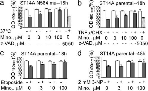
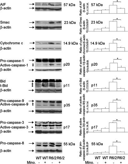
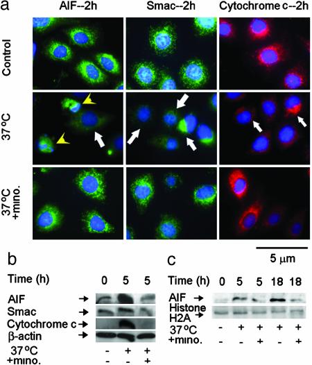
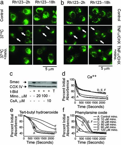
Minocycline is broadly protective in neurologic disease models featuring cell death and is being evaluated in clinical trials. We previously demonstrated that minocycline-mediated protection against caspase-dependent cell death related to its ability to prevent mitochondrial cytochrome c release. These results do not explain whether or how minocycline protects against caspase-independent cell death. Furthermore, there is no information on whether Smac/Diablo or apoptosis-inducing factor might play a role in chronic neurodegeneration. In a striatal cell model of Huntington's disease and in R6/2 mice, we demonstrate the association of cell death/disease progression with the recruitment of mitochondrial caspase-independent (apoptosis-inducing factor) and caspase-dependent (Smac/Diablo and cytochrome c) triggers. We show that minocycline is a drug that directly inhibits both caspase-independent and -dependent mitochondrial cell death pathways. Furthermore, this report demonstrates recruitment of Smac/Diablo and apoptosis-inducing factor in chronic neurodegeneration. Our results further delineate the mechanism by which minocycline mediates its remarkably broad neuroprotective effects.


Figures

Fig. 1.
Fig. 1.
Minocycline inhibition of striatal neuronal cell death. N584 mutant huntingtin-stable ST14A cells (a) and ST14A parental cells (b-d) were treated at the nonpermissive temperature of 37°C in SDM for 18 h (a), TNF-α (10 ng/ml)/CHX (10 μM) for 18 h (b), etoposide (50 μM) for 18 h (c), and 3-NP (2 mM) for 48 h (d) in the presence (3, 10, or 100 μM) or absence of minocycline. Cell death was evaluated by the 3-(4,5-dimethylthiazol-2-yl)-5-(3-carboxymethoxyphenyl)-2-(4-sulfophenyl)-2H-tetrazolium, inner salt assay and expressed as the percent of the control condition. The results are the mean ± SD of three independent experiments (*, P < 0.05). Empty bars indicate cells without apoptotic inducers. Solid bars indicate cells with apoptotic inducers. Hatched bars indicate cells with apoptotic inducers plus z-VAD.fmk. Mino represents minocycline-treated cells.
Fig. 5.
Fig. 5.
Minocycline inhibits AIF, Smac, and cytochrome c release, caspase activation, and Bid cleavage in R6/2 mice. Cytosolic fractions from 10.5-week-old R6/2 mice or wild-type mice treated with i.p. injections of saline or minocycline were analyzed by Western blot with AIF, Smac, and cytochrome c antibodies. Brain lysates from above mice were separated by SDS/PAGE and probed with caspase-1, -9, -3, and -8 and Bid antibodies. The same blot was reprobed with a β-actin antibody. Densitometry was performed to quantify each lane (n = 3 mice per condition, *, P < 0.05).
Fig. 2.
Fig. 2.
Minocycline inhibits the release of AIF, Smac, and cytochrome c in mutant huntingtin-expressing stable ST14A cells. (a) Mutant huntingtin ST14A cells were shifted at the nonpermissive temperature of 37°C in SDM with or without 10 μM minocycline for 2 h. The cells were fixed and stained with antibodies to AIF, Smac, or cytochrome c. Nuclei were visualized with Hoechst 33342 staining. Arrows indicate the release of AIF, Smac, and cytochrome c. Arrowhead indicates AIF translocation from mitochondria to nuclei by the overlap of AIF and Hoechst 33342 nuclear staining. (Bar = 5 μm.) (b and c) Mutant huntingtin ST14A cells were treated with or without shifting to the nonpermissive temperature for the indicated times. Cytosolic components (b) or nuclear extracts (c) were obtained, and samples (50 μg) were analyzed by Western blot with AIF, Smac, or cytochrome c antibodies. β-Actin was used as a loading control. A single blot, which was stripped, was used for all of the Western blots (b). Histone H2A was used as a nuclear extract loading control (c). The blot is representative of three independent experiments.
Fig. 3.
Fig. 3.
Minocycline inhibits caspase-8, -1, -9, and -3 activation and Bid cleavage. Mutant huntingtin stable ST14A cells were shifted to the nonpermissive temperature with or without 10 μM minocycline. Cells were extracted for immunoblotting (50 μg per lane) with anticaspase-8, -1, -9, -3 antibodies or anti-Bid antibody. The same blot was reprobed with β-actin antibody and used as a control for equal loading. The blots are representative of three independent experiments.
Fig. 4.
Fig. 4.
Minocycline inhibits dissipation of Δψm, release of Smac, and induction of PT. (a and b) Mutant huntingtin-expressing ST14 A cells were shifted at the nonpermissive temperature of 37°C in SDM (a), or ST14 A cells were treated with 10 ng/ml TNF-α/10 μM CHX (b) with or without 10 μM minocycline for 2 and 18 h. Cells were stained directly with 2 μM rhodamine 123. Arrows show dissipation of Δψm. (c) Purified mouse brain mitochondria were incubated with minocycline or cyclosporin A. Smac release was induced by tBid and inhibited by addition of minocycline or cyclosporin A. T, total mitochondrial Smac; CsA, cyclosporin A. With the mitochondrial pellets, cytochrome c oxidase subunit IV (COX IV) was used as a loading control. (d-f) Minocycline has distinct mechanisms of action against oxidative and nonoxidative PT inducers. Representative traces from a minimum of three experiments on the effects of minocycline on PT induction. Inducers: 50 μM Ca/Pi (d); 1 mM tert-butyl hydroperoxide 2 μMCa2+ (e); 50 μM phenylarsine oxide (f). PT induction was studied in liver mitochondria isolated from 4- to 6-month-old male Fischer 344 rats. Data for all traces in a single panel were collected at the same time from the same mitochondrial preparation.

References

    1. Yrjanheikki, J., Keinanen, R., Pellikka, M., Hokfelt, T. & Koistinaho, J. (1998) Proc. Natl. Acad. Sci. USA 95, 15769-15774. - PMC - PubMed
    1. Chen, M., Ona, V. O., Li, M., Ferrante, R. J., Fink, K. B., Zhu, S., Bian, J., Guo, L., Farrell, L. A., Hersch, S. M., et al. (2000) Nat. Med. 6, 797-801. - PubMed
    1. Zhu, S., Stavrovskaya, I. G., Drozda, M., Kim, B. Y., Ona, V., Li, M., Sarang, S., Liu, A. S., Hartley, D. M., Wu, du C., et al. (2002) Nature 417, 74-78. - PubMed
    1. Sanchez Mejia, R. O., Ona, V. O., Li, M. & Friedlander, R. M. (2001). Neurosurgery 48, 1393-1399; discussion, 1399-1401. - PubMed
    1. Popovic, N., Schubart, A., Goetz, B. D., Zhang, S. C., Linington, C. & Duncan, I. D. (2002) Ann. Neurol. 51, 215-223. - PubMed

Publication types