. 2003 Sep 2;100(18):10382-7.
doi: 10.1073/pnas.1832170100. Epub 2003 Aug 20.

Ian4 is required for mitochondrial integrity and T cell survival

Affiliations

Ian4 is required for mitochondrial integrity and T cell survival

Malini Pandarpurkar et al. Proc Natl Acad Sci U S A. .

Abstract

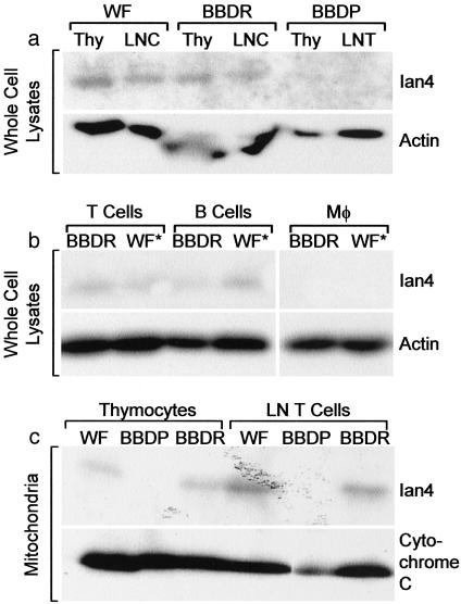
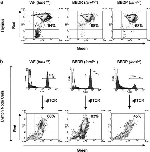
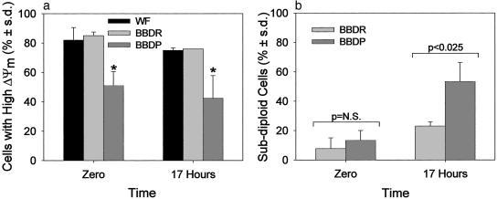
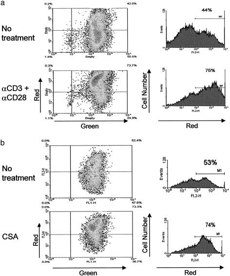
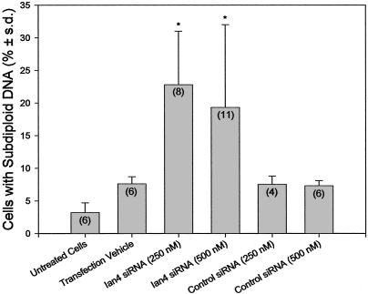
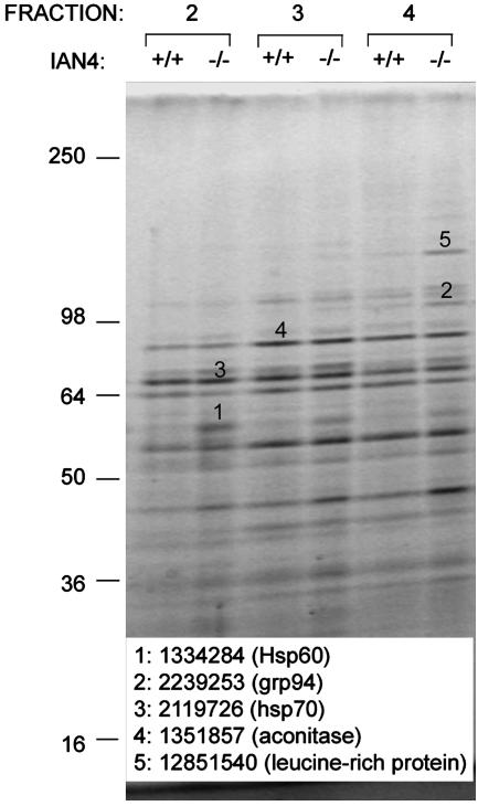
Apoptosis is a regulated cell death program controlled by extrinsic and intrinsic signaling pathways. The intrinsic pathway involves stress signals that activate pro-apoptotic members of the Bcl-2 family, inducing permeabilization of mitochondria and release of apoptogenic factors. These proteins localize to the outer mitochondrial membrane. Ian4, a mitochondrial outer membrane protein with GTP-binding activity, is normally present in thymocytes, T cells, and B cells. We and others have recently discovered that a mutation in the rat Ian4 gene results in severe T cell lymphopenia that is associated with the expression of autoimmune diabetes. The mechanism by which Ian4 controls T cell homeostasis is unknown. Here we show that the absence of Ian4 in T cells causes mitochondrial dysfunction, increased mitochondrial levels of stress-inducible chaperonins and a leucine-rich protein, and T cell-specific spontaneous apoptosis. T cell activation and caspase 8 inhibition both prevented apoptosis, whereas transfection of T cells with Ian4-specific small interfering RNA recapitulated the apoptotic phenotype. The findings establish Ian4 as a tissue-specific regulator of mitochondrial integrity.


Figures

Fig. 1.
Fig. 1.
Western analyses of Ian4 protein. (a) Thymocyte (Thy) and lymph node cell (LNC) lysates from Ian4+/+ BBDR and WF and Ian4-/- BBDP rats. (b) Lymph node T cell, splenic B cell, and macrophage lysates from Ian4+/+ BBDR and WF.ART2a (WF*, see ref. 9) rats. (c) Purified mitochondrial lysates from T cells and thymocytes of Ian4+/+ BBDR and WF and Ian4-/- BBDP rats. Actin and cytochrome c were used as loading controls.
Fig. 2.
Fig. 2.
Mitochondrial membrane potential (Δψm). (a) The Δψm of thymocytes from Ian4+/+ (WF and BBDR) and Ian4-/- (BBDP) rats; shown are the relative percentages of cells fluorescing red (vertical axis, high Δψm) and green (horizontal axis, low Δψm). The majority of thymocytes from both Ian4+/+ and Ian4-/- rats display high Δψm. Data are representative of two independent experiments. (b) The Δψm of lymph node T cells. Horizontal bars in histograms indicate gates used to identify αβTCR+ cells. The T lymphopenia of BBDP rats is evident. The dot plots show that the percentage of T cells with high Δψm was lower in Ian4-/- than in Ian4+/+ rats. Data are representative of three independent experiments; the complete dataset is shown in Fig. 3a. Control incubations with the mitochondrial uncoupler carbonylcyanide m-chlorophenylhydrazone uniformly reduced the percentage of T cells with high Δψm to ≈4% (data not shown).
Fig. 3.
Fig. 3.
Kinetics of Δψm and DNA fragmentation. (a) Peripheral T lymphocytes from Ian4+/+ (WF and BBDR) rats and Ian4-/- (BBDP) rats were assayed for Δψm at time of isolation (time 0) and after 17 h of culture. Shown are results of three independent experiments analyzed by ANOVA. *, P < 0.003 at time 0 and P < 0.006 at 17 h vs. BBDR and WF. (b) The percentage of cells with subdiploid DNA in peripheral T lymphocytes from BBDR and BBDP rats was determined by propidium iodide staining and flow microfluorometry when isolated (time 0) and after 17 h of culture. Shown are results of three independent experiments.
Fig. 4.
Fig. 4.
Rescue of mitochondrial membrane potential. Ian4-/- BBDP T cells were incubated for 17 h without or with anti-CD3 plus anti-CD28 mAbs (a) or for 30 min without or with cyclosporin A (CSA) (b). Gates used to identify the percentage of cells with high Δψm are indicated by horizontal bars. Data are representative of three independent experiments. Both activation and cyclosporin A rescued Ian4-/- lymphocytes from loss of Δψm.
Fig. 5.
Fig. 5.
siRNA analyses. Transfection of Ian4-specific siRNA increased T cell apoptosis. Bars depict the percentage of cells with subdiploid DNA after 48 h of treatment. *, Statistically similar to each other and P < 0.025 vs. each of the other four groups. The number of independent measurements is shown in parentheses. Two other Ian4 siRNAs were less effective (data not shown).
Fig. 6.
Fig. 6.
Proteomic analyses. Purified mitochondrial proteins from Ian4+/+ WF and Ian4-/- BBDP rat T cells resolved by SDS/PAGE. Bands 1, 2, and 5 were differentially expressed in Ian4+/+ vs. Ian4-/- T cells, excised, and identified by MALDI-TOF mass spectrometry. Not all differentially expressed bands were examined. Bands 3 and 4, which were similarly expressed, were excised to verify the purity of the mitochondrial isolation. Identities and gene identifiers for excised bands are indicated. Shown is one of two similar gels.

Similar articles

Cited by

References

    1. Zimmermann, K. C., Bonzon, C. & Green, D. R. (2001) Pharmacol. Ther. 92, 57-70. - PubMed
    1. Kroemer, G. & Reed, J. C. (2000) Nat. Med. 6, 513-519. - PubMed
    1. Vander Heiden, M. G., Chandel, N. S., Li, X. X., Schumacker, P. T., Colombini, M. & Thompson, C. B. (2000) Proc. Natl. Acad. Sci. USA 97, 4666-4671. - PMC - PubMed
    1. Daheron, L., Zenz, T., Siracusa, L. D., Brenner, C. & Calabretta, B. (2001) Nucleic Acids Res. 29, 1308-1316. - PMC - PubMed
    1. Hornum, L., Rømer, J. & Markholst, H. (2002) Diabetes 51, 1972-1979. - PubMed

Publication types

LinkOut - more resources