. 2003 Sep 1;31(17):5090-100.
doi: 10.1093/nar/gkg665.

Analysis of the DNA joining repertoire of Chlorella virus DNA ligase and a new crystal structure of the ligase-adenylate intermediate

Affiliations

Analysis of the DNA joining repertoire of Chlorella virus DNA ligase and a new crystal structure of the ligase-adenylate intermediate

Mark Odell et al. Nucleic Acids Res. .

Abstract

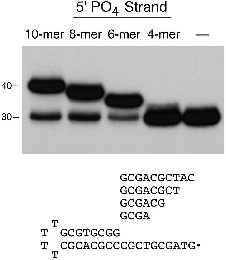
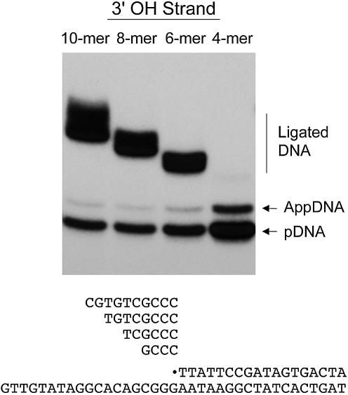
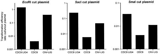
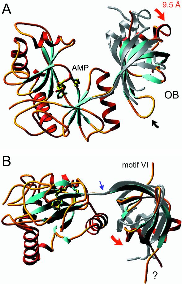
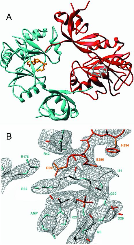
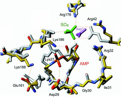
Chlorella virus DNA ligase is the smallest eukaryotic ATP-dependent DNA ligase known; it suffices for yeast cell growth in lieu of the essential yeast DNA ligase Cdc9. The Chlorella virus ligase-adenylate intermediate has an intrinsic nick sensing function and its DNA footprint extends 8-9 nt on the 3'-hydroxyl (3'-OH) side of the nick and 11-12 nt on the 5'-phosphate (5'-PO4) side. Here we establish the minimal length requirements for ligatable 3'-OH and 5'-PO4 strands at the nick (6 nt) and describe a new crystal structure of the ligase-adenylate in a state construed to reflect the configuration of the active site prior to nick recognition. Comparison with a previous structure of the ligase-adenylate bound to sulfate (a mimetic of the nick 5'-PO4) suggests how the positions and contacts of the active site components and the bound adenylate are remodeled by DNA binding. We find that the minimal Chlorella virus ligase is capable of catalyzing non-homologous end-joining reactions in vivo in yeast, a process normally executed by the structurally more complex cellular Lig4 enzyme. Our results suggest a model of ligase evolution in which: (i) a small 'pluripotent' ligase is the progenitor of the much larger ligases found presently in eukaryotic cells and (ii) gene duplications, variations within the core ligase structure and the fusion of new domains to the core structure (affording new protein-protein interactions) led to the compartmentalization of eukaryotic ligase function, i.e. by enhancing some components of the functional repertoire of the ancestral ligase while disabling others.


Figures

Figure 1
Figure 1
DNA length requirements on the 5′-PO4 side of the nick. Unlabeled 5′-phosphorylated oligonucleotides (10mer, 8mer, 6mer and 4mer) were annealed in 6-fold molar excess to a 5′ 32P-labeled 30mer tailed-hairpin strand to form the series of nicked substrates illustrated in the figure. Ligation reaction mixtures containing 25 nM radiolabeled DNA substrate as specified and 200 nM ligase were incubated for 10 min at 22°C. The radiolabeled products were resolved by denaturing gel electrophoresis and visualized by autoradiography. The positions of the input 30mer strand and the 40mer product of sealing the 10mer 5′-PO4 strand are indicated on the left.
Figure 2
Figure 2
DNA length requirements on the 3′-OH side of the nick. Unlabeled 3′-OH oligonucleotides (10mer, 8mer, 6mer and 4mer) were annealed in 6-fold molar excess to an unlabeled complementary template strand and a 5′ 32P-labeled 18mer strand to form the series of nicked substrates illustrated in the figure. Ligation reaction mixtures containing 25 nM radiolabeled DNA substrate as specified and 200 nM ligase were incubated for 10 min at 22°C. The radiolabeled products were resolved by denaturing gel electrophoresis and visualized by autoradiography. The positions of the input 5′ 32P-labeled 18mer (pDNA), the ligated DNA products and the DNA–adenylate intermediate (AppDNA) are indicated on the right.
Figure 3
Figure 3
Chlorella virus ligase catalyzes NHEJ reactions in vivo in yeast. NHEJ efficiency was determined using a linear plasmid transformation assay as described in Materials and Methods. The HIS3 plasmid was linearized by digestion with either EcoRI (left), SacI (middle) or SmaI (right). NHEJ efficiency was plotted on a log scale for the three yeast strains tested, which contained either the wild-type complement of yeast ligase genes (CDC9 and LIG4), CDC9 only (lig4Δ) or the Chlorella virus ligase gene only (cdc9Δ lig4Δ ChV-LIG).
Figure 4
Figure 4
Ligation of linear pUC19 DNA with cohesive overhangs. Reaction mixtures (10 µl) containing 50 mM Tris–HCl (pH 8.0), 5 mM DTT, 1 mM ATP, 0.2 µg (120 fmol) of pUC19 linearized with EcoR1 or SacI, and 0, 0.04, 0.4 or 4 ng of Chlorella virus DNA ligase (corresponding to ∼1, ∼10 or ∼100 fmol of enzyme) were incubated for 10 min at 22°C. The reaction products were analyzed by agarose gel electrophoresis in the presence of ethidium bromide. Uncut supercoiled pUC19 DNA was analyzed in parallel. A photograph of the stained gel under UV transillumination is shown. All lanes are from the same gel; the image was cropped to delete intervening lanes containing marker DNAs. The positions and sizes (in kilobase pairs) of linear DNA markers are indicated on the left. The species corresponding to linear dimer, nicked monomer circle, linear monomer and covalently closed monomer circle are indicated on the right.
Figure 5
Figure 5
New crystal structure of Chlorella virus ligase–adenylate. The ligase fold is shown with α helices in red and β strands in cyan. AMP is bound covalently to Lys27 in a pocket within the N-terminal nucleotidyl transferase domain. The C-terminal OB domain is connected to the nucleotidyl transferase domain by a flexible linker within motif V [denoted by the blue arrow in (B)]. The images in (A) and (B) show the positions of the OB domains of the current structure (the colored ribbon diagram) and the previous structure (the gray ribbon diagram) after superimposing the respective nucleotidyl transferase domains. Only the nucleotidyl transferase domain of the new structure is shown. The view in (A) is looking down onto the active site and putative DNA binding surface; this surface is located at the top of the ligase molecule in the view shown in (B). Movements of the OB domain are indicated by red arrows. A disordered surface loop of the OB domain is indicated by ‘?’ in (B).
Figure 6
Figure 6
Motif VI insinuates into the active site of a neighboring protomer. (A) Two symmetry-related ligase protomers are shown in cyan and red, respectively. The C-terminal peptide segments corresponding to motif VI project toward the active site of the neighboring protomer. (B) 2FoFc electron density map of the active site region contoured at the 1σ level. The map highlights the covalent lysyl–AMP adduct and the proximity to the adenylate of the C-terminal Asp297 side chain coming from motif VI of the neighboring protomer (colored in red).
Figure 7
Figure 7
Differences in the active sites of the two crystal structures of ligase–adenylate. The active sites of the old and new ligase crystal structures are shown in a superposition of the nucleotidyl transferase domains, with the old protein structure colored uniformly gray (except for the green sulfate ion) and the new structure colored according to atom identity (carbon in yellow, nitrogen in blue, oxygen in red, phosphorus in orange). The Asp297 carboxylate coming from the neighboring ligase protomer is shown in lavender (Asp*).

Similar articles

Cited by

References

    1. Doherty A.J. and Suh,S.W. (2000) Structural and mechanistic conservation in DNA ligases. Nucleic Acids Res., 28, 4051–4058. - PMC - PubMed
    1. Little J.W., Zimmerman,S.B., Oshinsky,C.K. and Gellert,M. (1967) Enzymatic joining of DNA strands: an enzyme–adenylate intermediate in the DPN-dependent DNA ligase reaction. Proc. Natl Acad. Sci. USA, 59, 2004–2011. - PMC - PubMed
    1. Weiss B., Thompson,A. and Richardson,C.C. (1968) Enzymatic breakage and rejoining of deoxyribonucleic acid: properties of the enzyme–adenylate intermediate in the polynucleotide ligase reaction. J. Biol. Chem., 246, 4523–4530. - PubMed
    1. Gumport R.I. and Lehman,I.R. (1971) Structure of the DNA ligase–adenylate intermediate: lysine (epsilon-amino)-linked adenosine monophosphoramidate. Proc. Natl Acad. Sci. USA, 68, 2559–2563. - PMC - PubMed
    1. Tomkinson A.E., Totty,N.F., Ginsburg,M. and Lindahl,T. (1991) Location of the active site for enzyme–adenylate formation in DNA ligases. Proc. Natl Acad. Sci. USA, 88, 400–404. - PMC - PubMed

Publication types

MeSH terms

Associated data