. 2003 Sep 1;31(17):5108-21.
doi: 10.1093/nar/gkg680.

3DNA: a software package for the analysis, rebuilding and visualization of three-dimensional nucleic acid structures

Affiliations

3DNA: a software package for the analysis, rebuilding and visualization of three-dimensional nucleic acid structures

Xiang-Jun Lu et al. Nucleic Acids Res. .

Abstract

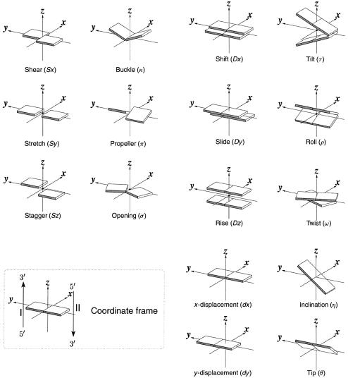
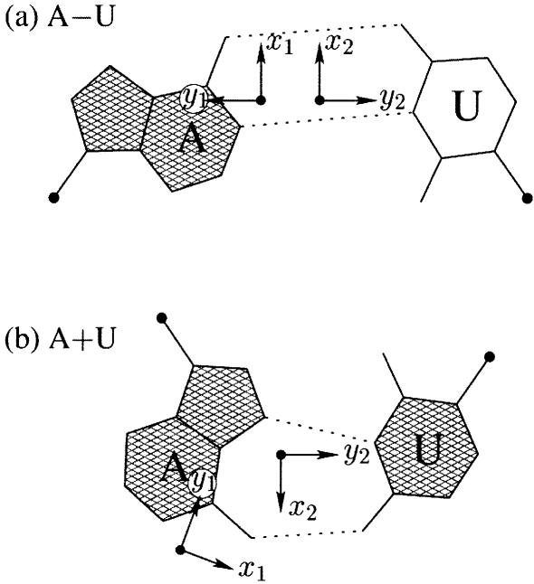
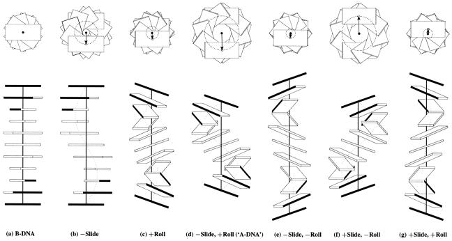
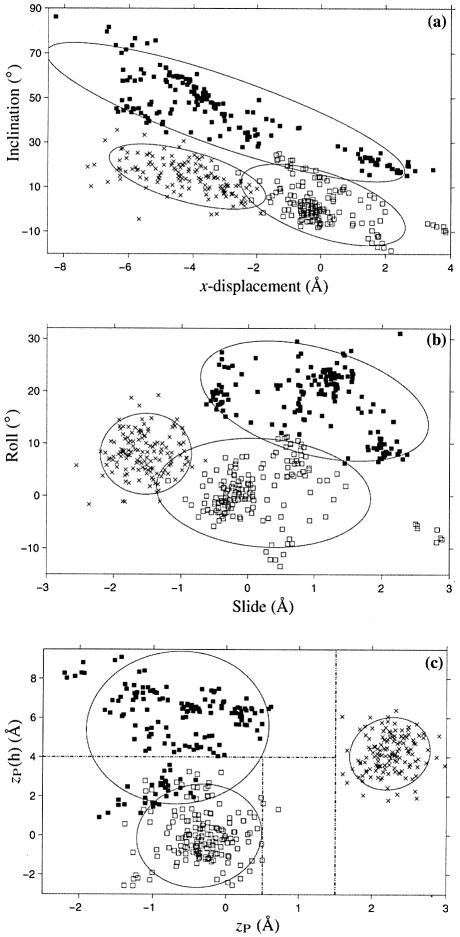
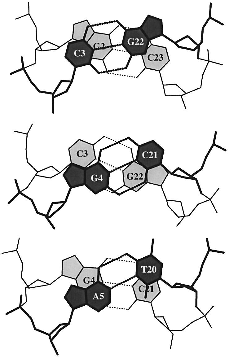
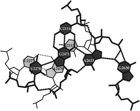
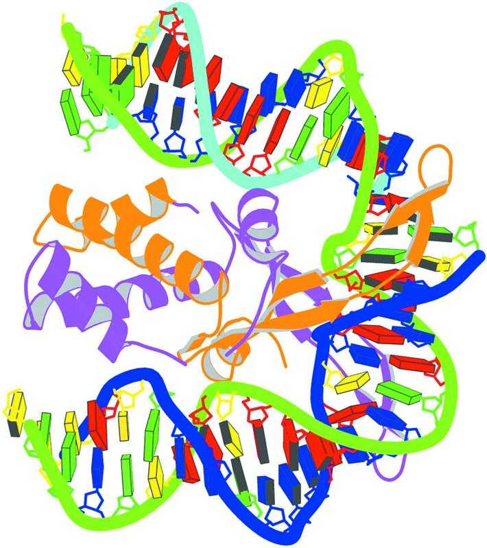
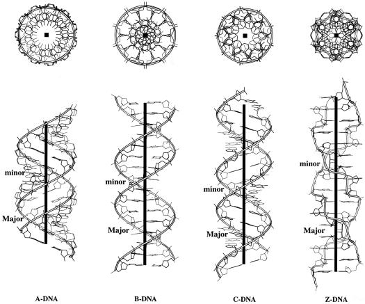
We present a comprehensive software package, 3DNA, for the analysis, reconstruction and visualization of three-dimensional nucleic acid structures. Starting from a coordinate file in Protein Data Bank (PDB) format, 3DNA can handle antiparallel and parallel double helices, single-stranded structures, triplexes, quadruplexes and other complex tertiary folding motifs found in both DNA and RNA structures. The analysis routines identify and categorize all base interactions and classify the double helical character of appropriate base pair steps. The program makes use of a recently recommended reference frame for the description of nucleic acid base pair geometry and a rigorous matrix-based scheme to calculate local conformational parameters and rebuild the structure from these parameters. The rebuilding routines produce rectangular block representations of nucleic acids as well as full atomic models with the sugar-phosphate backbone and publication quality 'standardized' base stacking diagrams. Utilities are provided to locate the base pairs and helical regions in a structure and to reorient structures for effective visualization. Regular helical models based on X-ray diffraction measurements of various repeating sequences can also be generated within the program.


Figures

Figure 1
Figure 1
Pictorial definitions of rigid body parameters used to describe the geometry of complementary (or non-complementary) base pairs and sequential base pair steps (19). The base pair reference frame (lower left) is constructed such that the x-axis points away from the (shaded) minor groove edge of a base or base pair and the y-axis points toward the sequence strand (I). The relative position and orientation of successive base pair planes are described with respect to both a dimer reference frame (upper right) and a local helical frame (lower right). Images illustrate positive values of the designated parameters. For illustration purposes, helical twist (Ωh) is the same as Twist (ω), formerly denoted by Ω (19,20) and helical rise (h) is the same as Rise (Dz).
Figure 2
Figure 2
Antiparallel and parallel combinations of adenine (A) and uracil (U) base pair ‘faces’: (a) the antiparallel Watson–Crick A–U pair with opposing faces (shaded versus unshaded) and a 1.5 Å Stretch introduced to separate the two base reference frames; (b) the parallel Hoogsteen A+U pair with base pair faces of the same sense. Black dots on bases denote the C1′ atoms on the attached sugars.
Figure 3
Figure 3
Large Shear of the G–U wobble base pair influences the calculated but not the ‘observed’ Twist. The 3DNA numerical values of Twist, 20° (top) and 43° (bottom), differ from the visualization of nearly equivalent Twist suggested by the angle between successive C1′···C1′ vectors (finely dotted lines). Illustrated dimer steps flank the G(8)–U(12) base pair in the crystal structure of the acceptor stem of E.coli tRNAAsp (NDB_ID: ar0019) (60).
Figure 4
Figure 4
Influence of non-zero Slide and Roll at sequential dimer steps on overall DNA helical conformation. Images generated with 3DNA building upon the principles of Calladine and Drew (42). The radii of the (dashed) circles in the upper row of images, defined by the loci of points from the helical axes (filled circles) to the base pair origin (open circles), correspond to the x-displacement. The values of dimeric Twist are adjusted, following equation 4, to keep the helical twist angle at 36°. The A-like model is highlighted in quotes to emphasize that the structure contains 10 rather than 11 residues per turn.
Figure 5
Figure 5
Scatter plots of selected conformational parameters showing the differences among A (× symbol), B (open squares) and TA DNA (filled squares) dinucleotide steps: (a) helical inclination and x-displacement; (b) dimer step Roll and Slide; (c) projected phosphorus positions, zP and zP(h). The contours correspond to ‘energies’ of 2kBT, i.e.‘2Δθ’ ellipses (17). Dashed lines in (c) illustrate the criteria used in 3DNA to distinguish the three helical forms (see text for details).
Figure 6
Figure 6
‘Standardized’ base stacking diagrams of three consecutive dimer steps of the 1.4 Å B DNA structure, d(CGCGAATTCGCG)2 (NDB_ID: bdl084) (87).
Figure 7
Figure 7
Stacking diagram of a U·G·C·A·G base pentad and a U·A·U triad in the refined structure of the large 50S ribosomal subunit (NDB_ID: rr0033) (3).
Figure 8
Figure 8
Schematic image of the IHF–DNA complex (NDB_ID: pdt040) (91) shown in the most extended view and color coded according to chain identity and DNA residue type, with the minor groove edges of base pairs shaded.
Figure 9
Figure 9
Top and side views illustrating the characteristic features of regular helical structures of A, B, C and Z DNA deduced from representative X-ray fiber diffraction models (44,46). Ribbons trace the progression of the backbone defined by the phosphorus atoms and the heavy black lines (boxes) represent the helical axes.

Similar articles

Cited by

References

    1. Watson J.D. and Crick,F.H.C. (1953) Genetical implications of the structure of deoxyribonucleic acid. Nature, 171, 964–967. - PubMed
    1. Saenger W. (1984) Principles of Nucleic Acid Structure. Springer-Verlag, New York, NY, Ch. 6.
    1. Klein D.J., Schmeing,T.M., Moore,P.B. and Steitz,T.A. (2001) The kink-turn: a new RNA secondary structure motif. EMBO J., 20, 4214–4221. - PMC - PubMed
    1. Kang C.H., Zhang,X., Ratliff,R., Moyzis,R. and Rich,A. (1992) Crystal structure of four-stranded Oxytricha telomeric DNA. Nature, 356, 126–131. - PubMed
    1. Smith F.W. and Feigon,J. (1992) Quadruplex structure of Oxytricha telomeric DNA oligonucleotides. Nature, 356, 164–168. - PubMed

Publication types