

Quantitation of intracellular NAD(P)H can monitor an imbalance of DNA single strand break repair in base excision repair deficient cells in real time
- PMID: 12930978
- PMCID: PMC212824
- DOI: 10.1093/nar/gng105
Quantitation of intracellular NAD(P)H can monitor an imbalance of DNA single strand break repair in base excision repair deficient cells in real time
Abstract
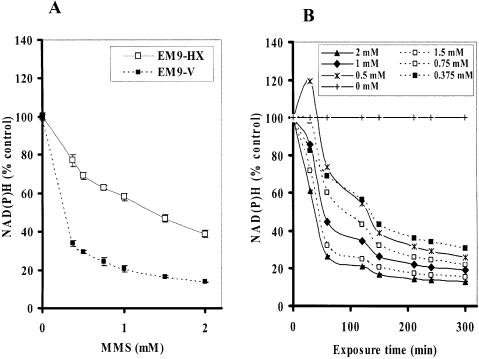
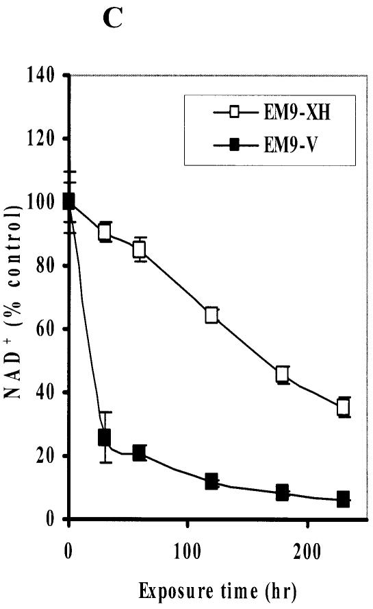
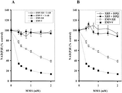
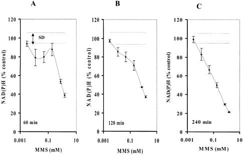
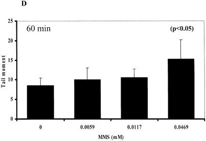
DNA single strand breaks (SSBs) are one of the most frequent DNA lesions in genomic DNA generated either by oxidative stress or during the base excision repair pathways. Here we established a new real-time assay to assess an imbalance of DNA SSB repair by indirectly measuring PARP-1 activation through the depletion of intracellular NAD(P)H. A water-soluble tetrazolium salt is used to monitor the amount of NAD(P)H in living cells through its reduction to a yellow colored water-soluble formazan dye. While this assay is not a direct method, it does not require DNA extraction or alkaline treatment, both of which could potentially cause an artifactual induction of SSBs. In addition, it takes only 4 h and requires less than a half million cells to perform this measurement. Using this assay, we demonstrated that the dose- and time-dependent depletion of NAD(P)H in XRCC1-deficient CHO cells exposed to methyl methanesulfonate. This decrease was almost completely blocked by a PARP inhibitor. Furthermore, methyl methanesulfonate reduced NAD(P)H in PARP-1+/+ cells, whereas PARP-1-/- cells were more resistant to the decrease in NAD(P)H. These results indicate that the analysis of intracellular NAD(P)H level using water-soluble tetrazolium salt can assess an imbalance of SSB repair in living cells in real time.
Figures

Similar articles
-
2,3,7,8-Tetrachlorodibenzo-p-dioxin (TCDD) induces oxidative stress, DNA strand breaks, and poly(ADP-ribose) polymerase-1 activation in human breast carcinoma cell lines.Toxicol Lett. 2007 Aug;172(3):146-58. doi: 10.1016/j.toxlet.2007.06.003. Epub 2007 Jun 16. Toxicol Lett. 2007. PMID: 17669606
-
Induction of ROS formation, poly(ADP-ribose) polymerase-1 activation, and cell death by PCB126 and PCB153 in human T47D and MDA-MB-231 breast cancer cells.Chem Biol Interact. 2006 Aug 25;162(2):181-94. doi: 10.1016/j.cbi.2006.06.009. Epub 2006 Jul 1. Chem Biol Interact. 2006. PMID: 16884709
-
Preventing oxidation of cellular XRCC1 affects PARP-mediated DNA damage responses.DNA Repair (Amst). 2013 Sep;12(9):774-85. doi: 10.1016/j.dnarep.2013.06.004. Epub 2013 Jul 18. DNA Repair (Amst). 2013. PMID: 23871146 Free PMC article.
-
XRCC1 and DNA strand break repair.DNA Repair (Amst). 2003 Sep 18;2(9):955-69. doi: 10.1016/s1568-7864(03)00118-6. DNA Repair (Amst). 2003. PMID: 12967653 Review.
-
Poly(ADP-ribose) Polymerase (PARP) and PARP Inhibitors: Mechanisms of Action and Role in Cardiovascular Disorders.Cardiovasc Toxicol. 2018 Dec;18(6):493-506. doi: 10.1007/s12012-018-9462-2. Cardiovasc Toxicol. 2018. PMID: 29968072 Review.
Cited by
-
SWI/SNF complexes are required for full activation of the DNA-damage response.Oncotarget. 2015 Jan 20;6(2):732-45. doi: 10.18632/oncotarget.2715. Oncotarget. 2015. PMID: 25544751 Free PMC article.
-
Selection for Improved Energy Use Efficiency and Drought Tolerance in Canola Results in Distinct Transcriptome and Epigenome Changes.Plant Physiol. 2015 Aug;168(4):1338-50. doi: 10.1104/pp.15.00155. Epub 2015 Jun 16. Plant Physiol. 2015. PMID: 26082400 Free PMC article.
-
Interplay between DNA polymerases beta and lambda in repair of oxidation DNA damage in chicken DT40 cells.DNA Repair (Amst). 2007 Jun 1;6(6):869-75. doi: 10.1016/j.dnarep.2007.01.011. Epub 2007 Mar 23. DNA Repair (Amst). 2007. PMID: 17363341 Free PMC article.
-
Chk2-dependent phosphorylation of XRCC1 in the DNA damage response promotes base excision repair.EMBO J. 2008 Dec 3;27(23):3140-50. doi: 10.1038/emboj.2008.229. Epub 2008 Oct 30. EMBO J. 2008. PMID: 18971944 Free PMC article.
-
Apoptosis induction by 13-acetoxyrolandrolide through the mitochondrial intrinsic pathway.Phytother Res. 2014 Jul;28(7):1045-53. doi: 10.1002/ptr.5091. Epub 2013 Dec 12. Phytother Res. 2014. PMID: 24338805 Free PMC article.
References
-
- Wood R.D. (1996) DNA repair in eukaryotes. Annu. Rev. Biochem., 65, 135–167. - PubMed
-
- Nakamura J., Walker,V.E., Upton,P.B., Chiang,S.-Y., Kow,Y.W. and Swenberg,J.A. (1998) Highly sensitive apurinic/apyrimidinic site assay can detect spontaneous and chemically induced depurination under physiological conditions. Cancer Res., 58, 222–225. - PubMed
-
- Lindahl T. (2000) Suppression of spontaneous mutagenesis in human cells by DNA base excision-repair. Mutat. Res., 462, 129–135. - PubMed
-
- Demple B. and Harrison,L. (1994) Repair of oxidative damage to DNA: enzymology and biology. Annu. Rev. Biochem., 63, 915–948. - PubMed
Publication types
MeSH terms
Substances
Grants and funding
LinkOut - more resources
Full Text Sources
Other Literature Sources
Miscellaneous
