

Structure of the Escherichia coli malate synthase G:pyruvate:acetyl-coenzyme A abortive ternary complex at 1.95 A resolution
- PMID: 12930982
- PMCID: PMC2323980
- DOI: 10.1110/ps.03174303
Structure of the Escherichia coli malate synthase G:pyruvate:acetyl-coenzyme A abortive ternary complex at 1.95 A resolution
Abstract
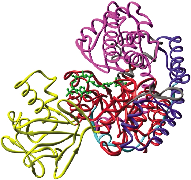
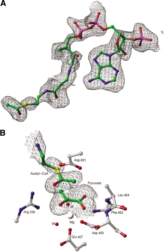
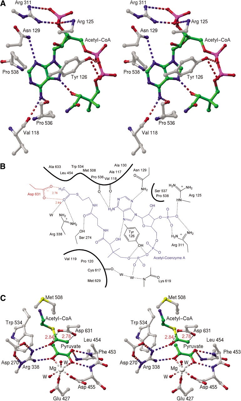
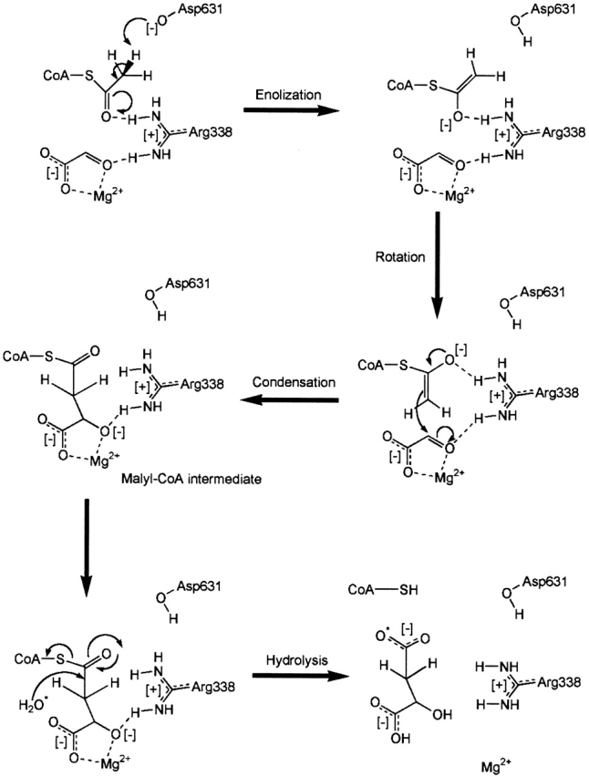
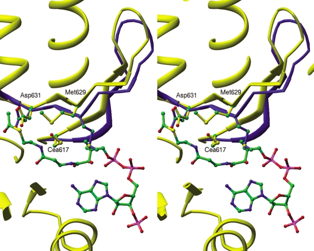
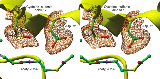
Malate synthase, an enzyme of the glyoxylate pathway, catalyzes the condensation and subsequent hydrolysis of acetyl-coenzyme A (acetyl-CoA) and glyoxylate to form malate and CoA. In the present study, we present the 1.95 A-resolution crystal structure of Escherichia coli malate synthase isoform G in complex with magnesium, pyruvate, and acetyl-CoA, and we compare it with previously determined structures of substrate and product complexes. The results reveal how the enzyme recognizes and activates the substrate acetyl-CoA, as well as conformational changes associated with substrate binding, which may be important for catalysis. On the basis of these results and mutagenesis of active site residues, Asp 631 and Arg 338 are proposed to act in concert to form the enolate anion of acetyl-CoA in the rate-limiting step. The highly conserved Cys 617, which is immediately adjacent to the presumed catalytic base Asp 631, appears to be oxidized to cysteine-sulfenic acid. This can explain earlier observations of the susceptibility of the enzyme to inactivation and aggregation upon X-ray irradiation and indicates that cysteine oxidation may play a role in redox regulation of malate synthase activity in vivo. There is mounting evidence that enzymes of the glyoxylate pathway are virulence factors in several pathogenic organisms, notably Mycobacterium tuberculosis and Candida albicans. The results described in this study add insight into the mechanism of catalysis and may be useful for the design of inhibitory compounds as possible antimicrobial agents.
Figures
References
-
- Appel, R.D., Baroch, A., and Hochstrasser, D.F. 1994. A new generation of information retrieval tools for biologists: The example of the ExPASy WWW server. Trends. Biochem. Sci. 19 258–261. - PubMed
-
- Bove, J., Martin, R.O., Ingraham, L.L., and Stumpf, P.K. 1959. Studies of the mechanism of action of the condensing enzyme. J. Biol. Chem. 234 999–1003. - PubMed
-
- Brandon, C. and Tooze, J. 1999. Introduction to protein structure. Garland Publishing, New York.
-
- Carson, M. 1997. Ribbons. Methods Enzymol. 277 493–505. - PubMed
Publication types
MeSH terms
Substances
Grants and funding
LinkOut - more resources
Full Text Sources
Molecular Biology Databases
