. 2003 Sep;12(9):1844-54.
doi: 10.1110/ps.03186703.

Solution structure of Pi4, a short four-disulfide-bridged scorpion toxin specific of potassium channels

Affiliations

Solution structure of Pi4, a short four-disulfide-bridged scorpion toxin specific of potassium channels

J Iñaki Guijarro et al. Protein Sci. 2003 Sep.

Erratum in

  • Protein Sci. 2003 Nov;12(11):2650. Possani Lourrival D [corrected to Possani Lourival D]

Abstract

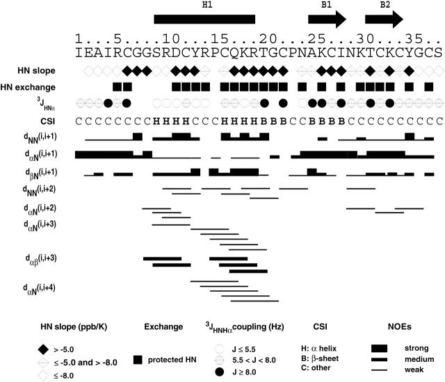
Pi4 is a short toxin found at very low abundance in the venom of Pandinus imperator scorpions. It is a potent blocker of K(+) channels. Like the other members of the alpha-KTX6 subfamily to which it belongs, it is cross-linked by four disulfide bonds. The synthetic analog (sPi4) and the natural toxin (nPi4) have been obtained by solid-phase synthesis or from scorpion venom, respectively. Analysis of two-dimensional (1)H NMR spectra of nPi4 and sPi4 indicates that both peptides have the same structure. Moreover, electrophysiological recordings of the blocking of Shaker B K(+) channels by sPi4 (K(D) = 8.5 nM) indicate that sPi4 has the same blocking activity of nPi4 (K(D) = 8.0 nM), previously described. The disulfide bonds have been independently determined by NMR and structure calculations, and by Edman-degradation/mass-spectrometry identification of peptides obtained by proteolysis of nPi4. Both approaches indicate that the pairing of the half-cystines is (6)C-(27)C, (12)C-(32)C, (16)C-(34)C, and (22)C-(37)C. The structure of the toxin has been determined by using 705 constraints derived from NMR data on sPi4. The structure, which is well defined, shows the characteristic alpha/beta scaffold of scorpion toxins. It is compared to the structure of the other alpha-KTX6 subfamily members and, in particular, to the structure of maurotoxin, which shows a different pattern of disulfide bridges despite its high degree of sequence identity (76%) with Pi4. The structure of Pi4 and the high amounts of synthetic peptide available, will enable the detailed analysis of the interaction of Pi4 with K(+) channels.


Figures

Figure 1.
Figure 1.
Sequence alignment of the αKTX6 subfamily. Cysteines are shown in blue. Solid lines represent disulfide-bridge pairings for standard (cyan), nonstandard (orange), and common to standard and nonstandard topologies (gray). MTX residues that have been mutated without any effect on its S–S bridge topology are labeled in green, and those that had an effect (change to standard topology) are colored in red (Fajloun et al. 2000c; Carlier et al. 2001). In magenta, two cysteine residues that have been mutated into α-aminobutyrate (Abu). The double Cys→Abu mutant adopts the standard arrangement of three-disulfide–bridged scorpion toxins (Fajloun et al. 2000b). Numbering corresponds to that of Pi4 and Pi7. The secondary structure of Pi4 as determined in this work is indicated by solid rectangles. (H indicates α-helix; B, β strand). The alignment was obtained with CLUSTAL W (Thompson et al. 1994).
Figure 2.
Figure 2.
Superposition of a region of the NOESY spectra of Pi4 extracted from scorpion venom (green) and obtained by chemical synthesis (blue). Spectra were acquired at 303 K by using a mixing time of 200 msec.
Figure 3.
Figure 3.
Pi4 disulfide-bridge assignment by NMR. Structures of Pi4 obtained with ARIA and CNS using no S–S constraint (magenta, structure a), ambiguous S–S constraints (cyan, structure b), and standard (blue, structure c) or nonstandard (red, structure d) disulfide topology are superimposed on backbone atoms between residues 4–37. The heavy atoms of side chains from Cys residues involved in S–S bridges that differ (black) or are common (green) to the standard and nonstandard S–S topologies are represented. The nonconvergent structure with nonstandard S–S arrangement is shifted upward for visualization purposes.
Figure 4.
Figure 4.
Summary of secondary structure NOE-related connectivities, amide exchange and temperature coefficient data, 3JHN-Hα coupling constants, and CSI (Wishart and Sykes 1994b) predictions. The secondary structure determined from the final structures is shown above the sequence: The helix H1 is represented by a rectangle, and the β-strands B1 and B2 by arrows.
Figure 5.
Figure 5.
Solution structure of Pi4. (A) Ribbon diagram of the best conformer (lowest total energy). (B) Stereoview of the calculated family of 10 conformers. Secondary structures are shown in blue and the rest of the backbone in red. Bonds between side chain heavy atoms of cysteine residues as well as disulfide bonds are displayed in green.
Figure 6.
Figure 6.
Synthetic Pi4 blocks Shaker B K+ channels with high affinity. (A, left) K+ currents through Shaker B channels evoked by 30-msec pulses from −30 to 60 mV. (Center) Residual K+ currents in the presence of 50 nM sPi4 in the external solution. (Right) K+ current recovery after washing the cell with the control external solution. (B) I–V relationship of the traces in A. (C) Fraction blocked as a function of [sPi4]. Fraction blocked = 1−(I/I0), where I is the peak current in the presence of the indicated [sPi4], and I0 is the control current, like in panel A. (Inset) The double reciprocal plot. The line corresponds to the least-squares fit of the points (KD = 8.5 nM, r = 0.988). Holding potential = −90 mV. The internal solution was (in mM) 90 KF, 30 KCl, 2 MgCl2, 10 EGTA, and 10 HEPES-K (pH 7.2), whereas the external solution was (in mM) 145 NaCl, 10 CaCl2, and 10 HEPES-Na (pH 7.2).

Similar articles

Cited by

References

    1. Batista, C.V.F., Gómez-Lagunas, F., Lucas, S., and Possani, L.D. 2000. Tc1, from Tityus cambridgei, is the first member of a new subfamily of scorpion toxins that blocks K+-channels. FEBS Lett. 486 117–120. - PubMed
    1. Bax, A. and Davis, D.G. 1985. MLEV-17–based two-dimensional homonuclear magnetization transfer spectroscopy. J. Magn. Reson. 65 355–360.
    1. Blanc, E., Sabatier, J.M., Kharrat, R., Meunier, S., El Ayeb, M., Van Rietschoten, J., and Darbon, H. 1997. Solution structure of maurotoxin, a scorpion toxin from Scorpio maurus, with high affinity for voltage-gated potassium channels. Protein 29 321–333. - PubMed
    1. Boisbouvier, J., Blackledge, M., Sollier, A., and Marion, D. 2000. Simultaneous determination of disulphide bridge topology and three-dimensional structure using ambiguous intersulphur distance restraints: Possibilities and limitations. J. Biomol. NMR 16 197–208. - PubMed
    1. Bontems, F., Roumestand, C., Gilquin, B., Ménez, A., and Toma, F. 1991. Refined structure of charybdotoxin: Common motifs in scorpion toxins and insects defensins. Science 253 1521–1523. - PubMed

Publication types

Associated data

LinkOut - more resources