. 2003 Sep;12(9):1894-901.
doi: 10.1110/ps.03114903.

Calculating pKa values in enzyme active sites

Affiliations

Calculating pKa values in enzyme active sites

Jens Erik Nielsen et al. Protein Sci. 2003 Sep.

Abstract

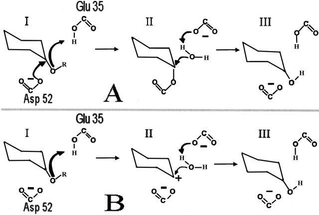
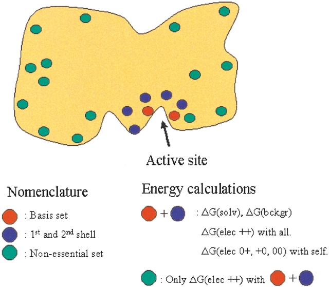
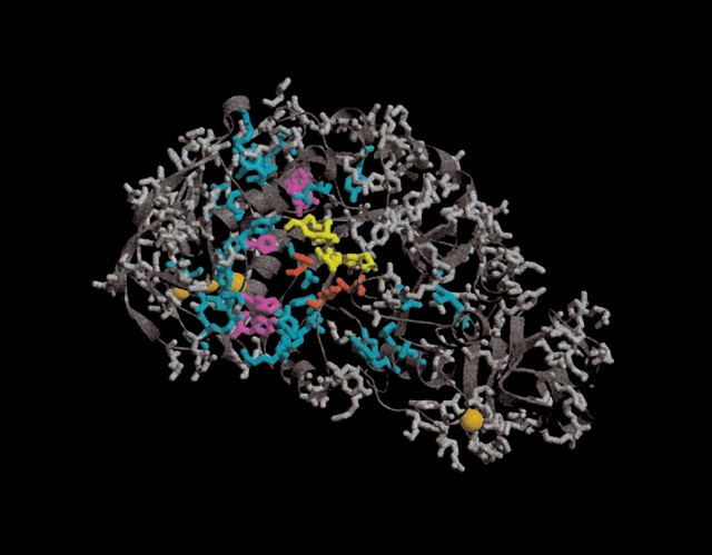
The ionization properties of the active-site residues in enzymes are of considerable interest in the study of the catalytic mechanisms of enzymes. Knowledge of these ionization constants (pKa values) often allows the researcher to identify the proton donor and the catalytic nucleophile in the reaction mechanism of the enzyme. Estimates of protein residue pKa values can be obtained by applying pKa calculation algorithms to protein X-ray structures. We show that pKa values accurate enough for identifying the proton donor in an enzyme active site can be calculated by considering in detail only the active-site residues and their immediate electrostatic interaction partners, thus allowing for a large decrease in calculation time. More specifically we omit the calculation of site-site interaction energies, and the calculation of desolvation and background interaction energies for a large number of pairs of titratable groups. The method presented here is well suited to be applied on a genomic scale, and can be implemented in most pKa calculation algorithms to give significant reductions in calculation time with little or no impact on the accuracy of the results. The work presented here has implications for the understanding of enzymes in general and for the design of novel biocatalysts.


Figures

Figure 1.
Figure 1.
The general catalytic mechanism for retaining glycosyl hydrolases (A), and the alternative mechanism for HEWL (B). (A) I: Protonation of the glycosidic oxygen by the proton donor (Glu 35) and attack on the glucose C1 by the nucleophile (Asp 52). Departure of the reducing end of the substrate. II: Activation of a water molecule, cleavage of C1-Asp 52 covalent bond. III: Regeneration of the initial protonation states. The residue numbering corresponds to that of HEWL. (B) The protonation of the glycosidic oxygen by Glu 35 results in the formation of an oxo-carbenium ion which is generated by an SN1 elimination of the R-OH group (I). The highly charged intermediate (II) is relaxed by reacting with a water molecule that neutralizes the positive charge on the substrate and regenerates the initial protonation state of Glu 35 (III).
Figure 2.
Figure 2.
The division of titratable groups used in the present pKa calculation scheme. Spheres indicate titratable groups. The figure shows a typical enzyme with most titratable groups at the surface and a higher concentration of titratable groups in and near the active site. The electrostatic energies calculated for each of the subsets are shown in the right text box. ΔG(elec ++) denotes charged-charged interaction energies, ΔG(elec 0+, +0, 00) denotes neutral-charged, charged-neutral, and neutral-neutral interaction energies.
Figure 3.
Figure 3.
Division of the titratable groups in Bacillus licheniformis α-amylase. Side chains are shown only for titratable groups. The three active-site residues Asp 231, Glu 261, and Asp 328 (BLA numbering) are colored red. The orange spheres represent inorganic ions present in the X-ray structure. Groups colored yellow are the additional residues that constitute the full subset when Ecutoff is 2.0 kT/e. Additional titratable groups that are included in the full subset when Ecutoff is lowered to 1.0 kT/e are colored magenta. The cyan groups are included when Ecutoff is lowered to 0.5 kT/e. Groups that always are in the nonessential set are colored gray.

References

    1. Alexov, E.G. and Gunner, M.R. 1997. Incorporating protein conformational flexibility into the calculation of pH-dependent protein properties. Biophys. J. 72 2075–2093. - PMC - PubMed
    1. Antosiewicz, J., McCammon, J.A., and Gilson, M.K. 1994. Prediction of pH-dependent properties of proteins. J. Mol. Biol. 238 415–436. - PubMed
    1. Baker, N.A., Sept, D., Joseph, S., Holst, M.J., and McCammon, J.A. 2001. Electrostatics of nanosystems: Application to microtubules and the ribosome. Proc. Natl. Acad. Sci. 98 10037–10041. - PMC - PubMed
    1. Bashford, D. and Karplus, M. 1990. pKa’s of ionizable groups in proteins: Atomic detail from a continuum electrostatic model. Biochemistry 29 10219–10225. - PubMed
    1. Berman, H.M., Westbrook, J., Feng, Z., Gilliland, G., Bhat, T.N., Weissig, H., Shindyalov, I.N., and Bourne, P.E. 2000. The Protein Data Bank. Nucleic Acids Res. 28 235–242. - PMC - PubMed

Publication types

LinkOut - more resources