. 2003 Sep;12(9):1980-90.
doi: 10.1110/ps.03166903.

Dissecting interdomain communication within cAPK regulatory subunit type IIbeta using enhanced amide hydrogen/deuterium exchange mass spectrometry (DXMS)

Affiliations

Dissecting interdomain communication within cAPK regulatory subunit type IIbeta using enhanced amide hydrogen/deuterium exchange mass spectrometry (DXMS)

Kerri M Zawadzki et al. Protein Sci. 2003 Sep.

Abstract

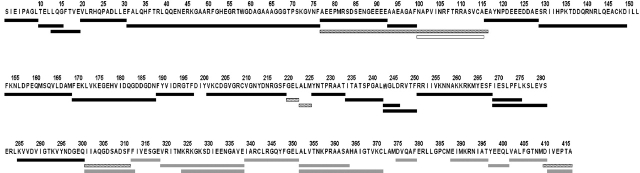
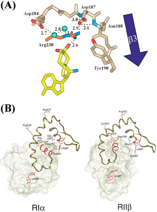
cAMP-dependent protein kinase (cAPK) is a heterotetramer containing a regulatory (R) subunit dimer bound to two catalytic (C) subunits and is involved in numerous cell signaling pathways. The C-subunit is activated allosterically when two cAMP molecules bind sequentially to the cAMP-binding domains, designated A and B (cAB-A and cAB-B, respectively). Each cAMP-binding domain contains a conserved Arg residue that is critical for high-affinity cAMP binding. Replacement of this Arg with Lys affects cAMP affinity, the structural integrity of the cAMP-binding domains, and cAPK activation. To better understand the local and long-range effects that the Arg-to-Lys mutation has on the dynamic properties of the R-subunit, the amide hydrogen/deuterium exchange in the RIIbeta subunit was probed by electrospray mass spectrometry. Mutant proteins containing the Arg-to-Lys substitution in either cAMP-binding domain were deuterated for various times and then, prior to mass spectrometry analysis, subjected to pepsin digestion to localize the deuterium incorporation. Mutation of this Arg in cAB-A (Arg230) causes an increase in amide hydrogen exchange throughout the mutated domain that is beyond the modest and localized effects of cAMP removal and is indicative of the importance of this Arg in domain organization. Mutation of Arg359 (cAB-B) leads to increased exchange in the adjacent cAB-A domain, particularly in the cAB-A domain C-helix that lies on top of the cAB-B domain and is believed to be functionally linked to the cAB-B domain. This interdomain communication appears to be a unidirectional pathway, as mutation of Arg230 in cAB-A does not effect dynamics of the cAB-B domain.


Figures

Figure 1.
Figure 1.
Overview of the R-subunits and the RIIβ phosphate binding cassettes (PBC). (A) (Top) Schematic diagram of R-subunit domain organization. Dimerization/docking domain is shown in brown; cAMP-binding domains A and B are shown in tan and in gray, respectively; and the pseudosubstrate inhibitor sequence is shown in yellow. (Bottom) Ribbon diagram of the cAMP-binding domains with the PBC-conserved Arg residues highlighted in orange. (B, C) Close-up diagram highlighting the network of interactions originating from Arg230 and Arg359 (B and C, respectively). Acidic and basic side-chain atoms are shown in red and blue, respectively. Hydrogen bonds are designated with dashed lines, and water molecules are shown in aqua. Residues labeled as, e.g., G220:N, indicate that the H-bond is directed to the amide group of G220.
Figure 2.
Figure 2.
Pepsin digestion map of R230K and R359K RIIβ. Black lines indicate peptides analyzed in all wild type, R230K, and R359K. Gray lines indicate peptides analyzed in wild type and R230K. Dashed lines indicate peptides analyzed in R230K and R359K. White lines indicate peptides analyzed in wild-type protein only.
Figure 3.
Figure 3.
Deuteration levels of RIIβ. Each horizontal grouping of bars represents, from top to bottom, cAMP-bound wild type, R230K, R359K, and cAMP-free wild type. Each data set contains six time points: 10-s, 30-s, 100-s, 300-s, 1000-s, and 3000-s on-exchange.
Figure 4.
Figure 4.
Difference in deuteration between Arg mutant and wild-type proteins mapped onto ribbon diagram of RIIβ residues 130–412. Residues showing increased deuteration upon mutation are in red, decreased deuteration upon mutation are in blue, and little change in deuteration are in gray. Arg230 (A) and Arg359 (B) are shown in green.
Figure 5.
Figure 5.
Number of deuterons incorporated as a function of time for various peptides in wild-type cAMP-free (open circles), wild-type cAMP-bound (solid circles), R230K (triangles), and R359K (squares) RIIβ. (A) Amide hydrogen exchange of dimerization/docking domain helix I residues 12–15 and 15–19. (B) Residues within cAMP-binding domain A with amide exchange that is altered upon Arg mutation. Ribbon diagram of the cA domain highlighting (red) residues 171–188 (β1,β2), 191–197 (β3,β4), 203–219 (β5), 228–233 (PBC), 236–242 (β7,β8), 245–246 (β8), 247–250 (αB), 253–268 (αC), and 271–281 (αC). Arg230 is shown in green, and the conserved Asp residues, which interact with Arg230, are shown in white. The plot for residues 236–242 is representative of the plots for 191–197, 203–219, 245–246, 247–250, and 228–233 is representative of 171–188. (C) Residues within cAMP-binding domain B whose amide exchange is altered upon Arg mutation. Ribbon diagram of the cAB-B domain highlighting residues 287–300 (αA,β1). Arg359 is shown in green; the conserved Asp residues, which interact with Arg359, are shown in white; and Arg381 is shown in purple.
Figure 6.
Figure 6.
(A) Diagram highlighting the hydrogen bond interactions of Asp187. In addition to the Asp187-Arg230 (orange)-cAMP (yellow) bridge, Asp187 also interacts with the backbone of β-sheet 3. Acidic residues are shown in red, and basic residues are shown in blue. Water molecules are shown in aqua. Hydrogen bond distances are given in angstroms. (B) RIa and RIIβ surface diagrams highlighting the contacts between the cAB-A domain C-helix (green ribbon) and the cAB-B domain (green). The cAB-B domain B-helix (red) stacks against the cAB-A domain PBC.
Figure 7.
Figure 7.
(A) Amino acid side-chain and backbone interactions connecting shells I and II in the R-subunit. Residues of shell I (PBC, gold), shell II (β2–β3 turn, blue; cAB-B domain αA, cranberry; and cAB-A domain αB and αC, silver) are displayed. Two numbers are listed for each amino acid; the first corresponds to RIα numbering, and the second corresponds to RIIβ numbering. Interactions conserved between RIα and RIIβ are shown with black dotted lines. Interactions unique to RIα are shown with solid blue lines, and interactions unique to RIIβ are shown with solid green lines. The residues that provide the primary isoform difference between the functional shells (Arg241/262, Lys240/261, and Asp267/288) are designated with a box. (B, C) Ribbon diagrams of RIα and RIIβ demonstrating the isoform-specific connection between the cAB-A domain PBC and C-helix.

Similar articles

Cited by

References

    1. Amieux, P.S., Howe, D.G., Knickerbocker, H., Lee, D.C., Su, T., Laslo, G.S., Idzerda, R.L., and McKnight, G.S. 2002. Increased basal PKA activity inhibits the formation of mesoderm-derived structures in the developing mouse embryo. J. Biol. Chem. 277 27294–27304. - PubMed
    1. Anand, G.S., Hughes, C.A., Jones, J.M., Taylor, S.S., and Komives, E.A. 2002. Amide H/2H exchange reveals communication between the cAMP and catalytic subunit-binding sites in the RIα subunit of protein kinase A. J. Mol. Biol. 323 377–386. - PubMed
    1. Andersen, M.D., Shaffer, J., Jennings, P.A., and Adams, J.A. 2001. Structural characterization of protein kinase A as a function of nucleotide binding: Hydrogen-deuterium exchange studies using matrix-assisted laser desorption ionization-time of flight mass spectrometry detection. J. Biol. Chem. 276 14204–14211. - PubMed
    1. Bai, Y., Milne, J.S., Mayne, L.C., and Englander, S.W. 1993. Primary structure effects on peptide group hydrogen exchange. Proteins 17 75–86. - PMC - PubMed
    1. Banky, P., Newlon, M.G., Roy, M., Morikis, D., Haste, N.M., Taylor, S.S., and Jennings, P.A. 2003. Unique dimeric surface topology mediates isoform-specific anchoring of PKA. J. Mol. Biol. (in press).

Publication types

MeSH terms

LinkOut - more resources