

Encoding specificity in plant calcium signalling: hot-spotting the ups and downs and waves
- PMID: 12933365
- PMCID: PMC4243675
- DOI: 10.1093/aob/mcg173
Encoding specificity in plant calcium signalling: hot-spotting the ups and downs and waves
Abstract
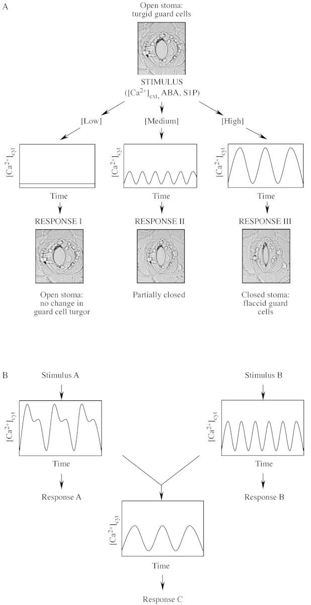
Calcium ions function as intracellular second messengers in regulating a plethora of cellular processes from acclimative stress responses to survival and programmed cell death. The generation of specificity in Ca2+ signals is dependent on influx and efflux from the extracellular milieu, cytosol and intracellular organelles. One aspect of plant Ca2+ signalling that is currently attracting a great deal of interest is how 'Ca2+-signatures', specific spatio-temporal changes in cytosolic-free Ca2+, encode the necessary information to bring about this range of physiological responses. Here, current information is reviewed on how Ca2+-signatures are generated in plant cells and how stimulus-specific information can be encoded in the form of Ca2+-signatures.
Figures
Similar articles
-
Shaping the calcium signature.New Phytol. 2009 Jan;181(2):275-294. doi: 10.1111/j.1469-8137.2008.02682.x. New Phytol. 2009. PMID: 19121028 Review.
-
Calcium in plants.Ann Bot. 2003 Oct;92(4):487-511. doi: 10.1093/aob/mcg164. Epub 2003 Aug 21. Ann Bot. 2003. PMID: 12933363 Free PMC article. Review.
-
Mitochondria as biosensors of calcium microdomains.Cell Calcium. 1999 Nov;26(5):193-9. doi: 10.1054/ceca.1999.0076. Cell Calcium. 1999. PMID: 10643557 Review.
-
[Extracellular Ca2+ signaling: first messenger in animals and plants].Yi Chuan. 2007 Mar;29(3):269-75. doi: 10.1360/yc-007-0269. Yi Chuan. 2007. PMID: 17369145 Review. Chinese.
-
Remodelling of Ca2+ transport in cancer: how it contributes to cancer hallmarks?Philos Trans R Soc Lond B Biol Sci. 2014 Feb 3;369(1638):20130097. doi: 10.1098/rstb.2013.0097. Print 2014 Mar 19. Philos Trans R Soc Lond B Biol Sci. 2014. PMID: 24493745 Free PMC article. Review.
Cited by
-
Respective contribution of CML8 and CML9, two arabidopsis calmodulin-like proteins, to plant stress responses.Plant Signal Behav. 2017 May 4;12(5):e1322246. doi: 10.1080/15592324.2017.1322246. Epub 2017 May 4. Plant Signal Behav. 2017. PMID: 28471263 Free PMC article.
-
A possible mechanism and sequence of events that lead to the Al3+-induced [Ca2+]cyt transients and inhibition of root growth.Plant Signal Behav. 2010 Jul;5(7):881-4. doi: 10.4161/psb.5.7.11973. Epub 2010 Jul 1. Plant Signal Behav. 2010. PMID: 20495379 Free PMC article.
-
Calcium efflux systems in stress signaling and adaptation in plants.Front Plant Sci. 2011 Dec 2;2:85. doi: 10.3389/fpls.2011.00085. eCollection 2011. Front Plant Sci. 2011. PMID: 22639615 Free PMC article.
-
Embryos assist morphogenesis of others through calcium and ATP signaling mechanisms in collective teratogen resistance.Nat Commun. 2024 Jan 17;15(1):535. doi: 10.1038/s41467-023-44522-2. Nat Commun. 2024. PMID: 38233424 Free PMC article.
-
Signal percolation through plants and the shape of the calcium signature.Plant Signal Behav. 2010 Apr;5(4):379-85. doi: 10.4161/psb.5.4.10717. Epub 2010 Apr 20. Plant Signal Behav. 2010. PMID: 20139732 Free PMC article.
References
-
- AllenGJ, Chu SP, Harrington CL, Schumacher K, Hoffmann T, Tang YY, Grill E, Schroeder JI.2001. A defined range of guard cell calcium oscillation parameters encodes stomatal movements. Nature 411: 1053–1057. - PubMed
-
- AllenGJ, Chu SP, Schumacher K, Shimazaki CT, Vafeados D, Kemper A, Hawke SD, Tallman G, Tsien RY, Harper JFet al.2000. Alteration of stimulus‐specific guard cell calcium oscillations and stomatal closing in Arabidopsis det3 mutants. Science 289: 2338–2342. - PubMed
-
- BerkowitzG, Zhang X, Mercier R, Leng Q, Lawton M.2000. Co‐expression of calcium‐dependent protein kinase with the inward rectified guard cell K+ channel KAT1 alters current parameters in Xenopus laevis oocytes. Plant and Cell Physiology 41: 785–790. - PubMed
-
- BerridgeMJ, Cobbold PH, Cuthbertson KSR.1988. Spatial and temporal aspects of cell signalling. Philosophical Transactions of the Royal Society of London Series B 320: 325–343. - PubMed
-
- BerridgeMJ, Lipp P, Bootman MD.2000. The versatility and universality of calcium signalling. Nature Reviews – Molecular Cell Biology 1: 11–21. - PubMed
Publication types
MeSH terms
Substances
LinkOut - more resources
Full Text Sources
Miscellaneous
