. 2003 Nov 14;278(46):45629-37.
doi: 10.1074/jbc.M303292200. Epub 2003 Aug 20.

Keratocyte phenotype mediates proteoglycan structure: a role for fibroblasts in corneal fibrosis

Affiliations

Keratocyte phenotype mediates proteoglycan structure: a role for fibroblasts in corneal fibrosis

James L Funderburgh et al. J Biol Chem. .

Abstract

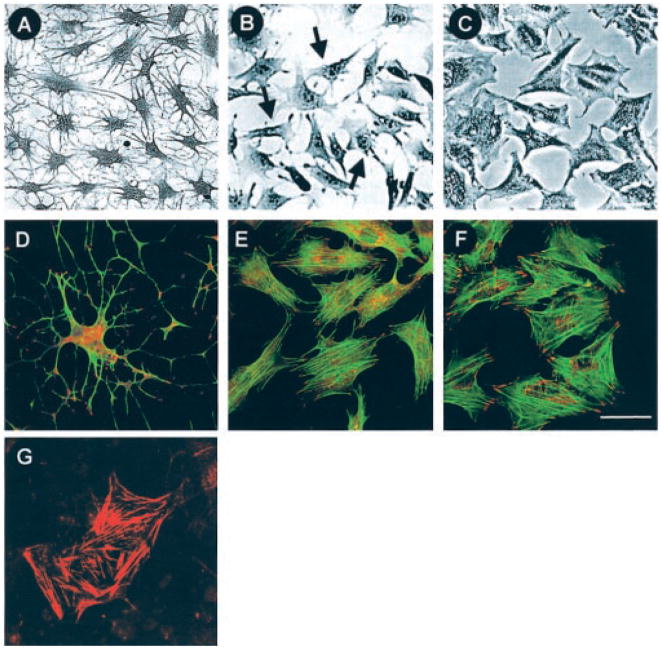
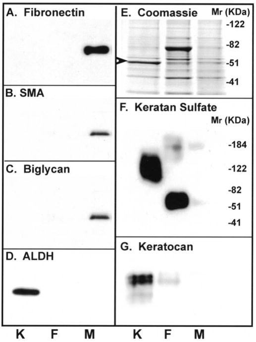
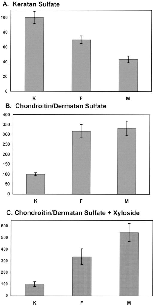
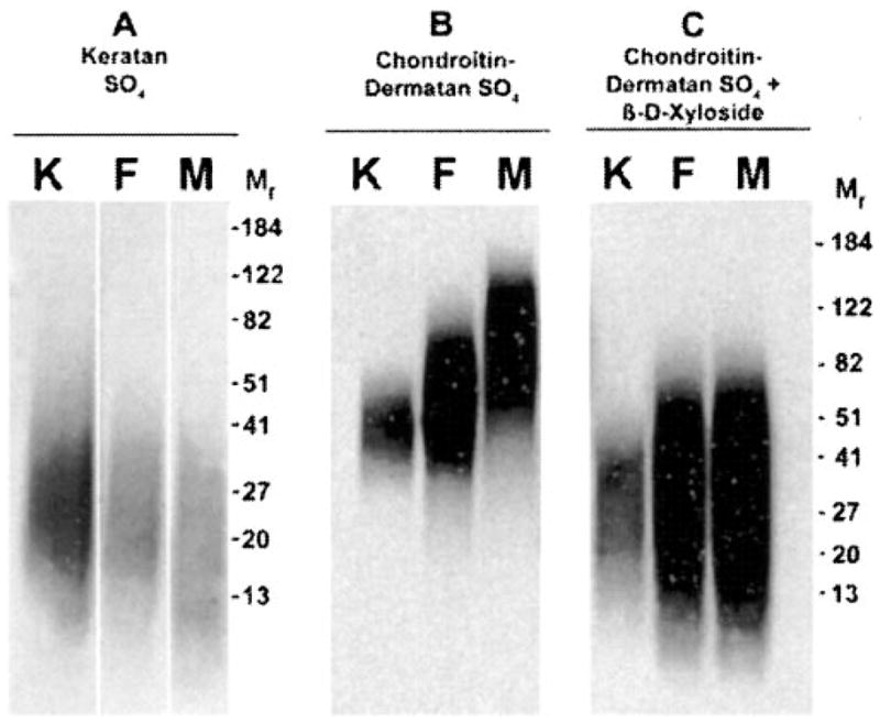
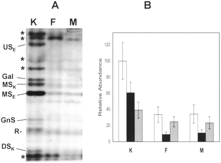
In pathological corneas, accumulation of fibrotic extracellular matrix is characterized by proteoglycans with altered glycosaminoglycans that contribute to the reduced transparency of scarred tissue. During wound healing, keratocytes in the corneal stroma transdifferentiate into fibroblasts and myofibroblasts. In this study, molecular markers were developed to identify keratocyte, fibroblast, and myofibroblast phenotypes in primary cultures of corneal stromal cells and the structure of glycosaminoglycans secreted by these cells was characterized. Quiescent primary keratocytes expressed abundant protein and mRNA for keratocan and aldehyde dehydrogenase class 3 and secreted proteoglycans containing macromolecular keratan sulfate. Expression of these marker compounds was reduced in fibroblasts and also in transforming growth factor-beta-induced myofibroblasts, which expressed high levels of alpha-smooth muscle actin, biglycan, and the extra domain A (EDA or EIIIA) form of cellular fibronectin. Collagen types I and III mRNAs were elevated in both fibroblasts and in myofibroblasts. Expression of these molecular markers clearly distinguishes the phenotypic states of stromal cells in vitro. Glycosaminoglycans secreted by fibroblasts and myofibroblasts were qualitatively similar to and differed from those of keratocytes. Chondroitin/dermatan sulfate abundance, chain length, and sulfation were increased as keratocytes became fibroblasts and myofibroblasts. Fluorophore-assisted carbohydrate electrophoresis analysis demonstrated increased N-acetylgalactosamine sulfation at both 4- and 6-carbons. Hyaluronan, absent in keratocytes, was secreted by fibroblasts and myofibroblasts. Keratan sulfate biosynthesis, chain length, and sulfation were significantly reduced in both fibroblasts and myofibroblasts. The qualitatively similar expression of glycosaminoglycans shared by fibroblasts and myofibroblasts suggests a role for fibroblasts in deposition of non-transparent fibrotic tissue in pathological corneas.


Figures

Fig. 1
Fig. 1. Morphology and cytoskeletal organization of cultured corneal keratocytes, fibroblasts, and myofibroblasts
Primary cultures of bovine keratocytes were established under conditions that either maintain the keratocyte phenotype or that initiate transdifferentiation to fibroblast or myofibroblastic phenotypes as described under “Experimental Procedures.” Micrographs A–C illustrate morphology of cells after staining with crystal violet. Panels D–F show cells stained with phalloidin (green) and anti-vinculin (red). In G, myofibroblasts were stained with antibodies to α-smooth muscle α actin. Keratocytes and fibroblasts were negative for α-smooth muscle α staining. White bars in C and F are 50 μm.
Fig. 2
Fig. 2. Immunoblotting of phenotypic marker proteins
A, cellular fibronectin, 200 kDa. B, α-smooth muscle α-actin (SMA), 54 kDa. C, biglycan (BGN), 49 kDa. D, ALDH, 54 kDa. E, total protein. F, keratan sulfate. G, keratocan, 50 kDa. All were detected in cell lysates (A, B, D, and E) or in conditioned media (C, F, and G) by immunoblotting after separation on SDS-PAGE as described under “Experimental Procedures.” Panel E shows a Coomassie Blue stain of a cell extract similar to that immunoblotted for ALDH in panel D. Arrow marks a prominent 54-kDa band from keratocytes corresponding to ALDH in the blot. Lane K, keratocytes; lane F, fibroblasts; and lane M, myofibroblasts.
Fig. 3
Fig. 3. Incorporation of [35S]sulfate into keratocyte glycosaminoglycans as a function of cell phenotype
Incorporation of [35S]sulfate into keratan sulfate and chondroitin/dermatan sulfate during an 18-h labeling period was determined by digestion of a purified proteoglycan fraction with keratanase II + endo-β-galactosidase (A) or chondroitinase ABC (B and C) as described under “Experimental Procedures.” Values are corrected for cell protein, and error bars represent the mean ± S.D. of assays on triplicate cultures. Values are normalized so that keratocyte = 100 in each assay. In C, cultures were labeled in the presence of 0.5 mM 4-nitrophenyl-β-D-xyloside.
Fig. 4
Fig. 4. Glycosaminoglycan chain size in cultured corneal cells
Proteoglycans from culture media after labeling with [35S]sulfate under conditions similar to Fig. 3 were separated into keratan sulfate proteoglycans using chondroitinase digestion (A) or chondroitin/dermatan sulfate using fractional alcohol precipitation (B and C), and then glycosaminoglycan chains were released by proteinase digestion as described under “Experimental Procedures.” Free glycosaminoglycan chains were separated SDS-PAGE and detected by autoradiography. Molecular size markers represent protein standards run in the same gels. In C, labeling was carried out in the presence of 0.5 mM 4-nitrophenyl-β-D-xyloside.
Fig. 5
Fig. 5. Analysis of hyaluronan and chondroitin/dermatan sulfate by FACE
A, unlabeled glycosaminoglycans were digested with chondroitinase ABC and fragments derivatized with 2-aminoacridone, separated by gel electrophoresis, and visualized by fluorescence as described under “Experimental Procedures.” Fragments were identified by co-electrophoresis with standards. Di-HA, hyaluronan-unsaturated disaccharide; Di-0S, chondroitin/dermatan-unsulfated unsaturated disaccharide; Di-4S, chondroitin/dermatan 4-O-sulfated unsaturated disaccharide; Di-6S, chondroitin/dermatan 6-O-sulfated unsaturated disaccharide; Di-diS, chondroitin/dermatan 4,6,-O-disulfated disaccharide. B, relative abundance of chondroitin/dermatan fragments was determined by quantitative image analysis of fluorescent gels similar to those in A. The total chondroitin/dermatan fragments in each lane was normalized to 100. Error bars show the mean ± S.D. of analyses of triplicate cultures. White bars, Di-0S; black bars, Di-6S; gray bars; Di-4S. C, hyaluronan fragments were calculated as in B as a percentage of chondroitin/dermatan sulfate in the same sample.
Fig. 6
Fig. 6. Analysis of keratan sulfate by FACE
A, unlabeled keratan sulfate from keratocyte cultures was digested with a mixture of keratanase II and endo-β-galactosidase. Fragments were derivatized with 2-aminoacridone, separated by gel electrophoresis, and detected by fluorescence as described under “Experimental Procedures.” Marked bands representing >90% of the labeled products were used for quantitation. Bands were identified by co-electrophoresis with commercial standards (Gal, GnSO4) or with fragments of purified corneal keratan sulfate characterized as described under “Experimental Procedures.” MSE and MSK, monosulfated fragments produced by endo-β-galactosidase (GnSO4-Gal) and keratanase II (Gal-GnSO4); USE, unsulfated endo-β-galactosidase disaccharide (Gn-Gal); DSK, disulfated keratanase II disaccharide (GalSO4-GnSO4); R, nonspecific reagent band. Bands marked with asterisk are keratan sulfate-derived components of non-determined structure. B, quantification of keratan sulfate bands in keratocyte, fibroblasts, and myofibroblasts. Triplicate samples similar to that in Fig. 6A were analyzed for abundance of the 11 bands marked in 6A. White bars show the sum of all of the components (excluding R). Black bars show sums of identified components of keratan sulfate chain elongation, and gray bars show unidentified fragments marked asterisk in A. The total for keratocytes (K) was set to 100.

Similar articles

Cited by

References

    1. Chakravarti S, Petroll WM, Hassell JR, Jester JV, Lass JH, Paul J, Birk DE. Invest Ophthalmol Visual Sci. 2000;41:3365–3373. - PMC - PubMed
    1. Funderburgh JL, Caterson B, Conrad GW. J Biol Chem. 1987;262:11634–11640. - PubMed
    1. Hassell JR, Cintron C, Kublin C, Newsome DA. Arch Biochem Biophys. 1983;222:362–369. - PubMed
    1. Hassell JR, Newsome DA, Krachmer JH, Rodrigues MM. Proc Natl Acad Sci U S A. 1980;77:3705–3709. - PMC - PubMed
    1. Nakazawa K, Hassell JR, Hascall VC, Lohmander LS, Newsome DA, Krachmer J. J Biol Chem. 1984;259:13751–13757. - PubMed

Publication types

MeSH terms

Associated data