. 2003 Sep;71(9):4891-900.
doi: 10.1128/IAI.71.9.4891-4900.2003.

Increased mortality and dysregulated cytokine production in tumor necrosis factor receptor 1-deficient mice following systemic Klebsiella pneumoniae infection

Affiliations

Increased mortality and dysregulated cytokine production in tumor necrosis factor receptor 1-deficient mice following systemic Klebsiella pneumoniae infection

Thomas A Moore et al. Infect Immun. 2003 Sep.

Abstract

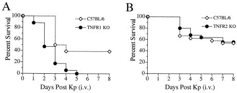
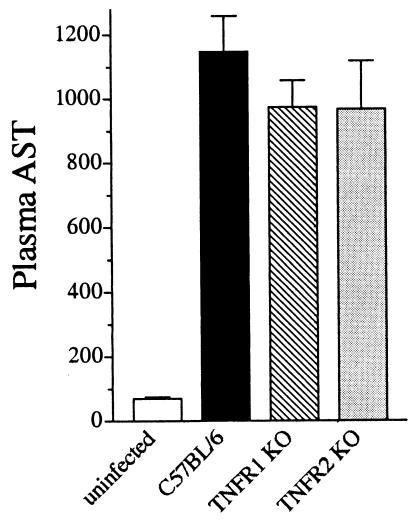
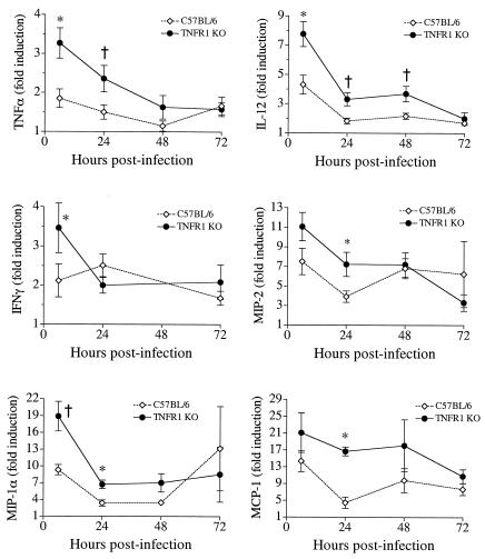
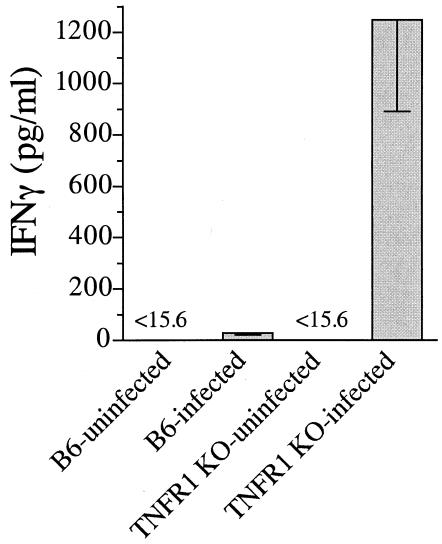
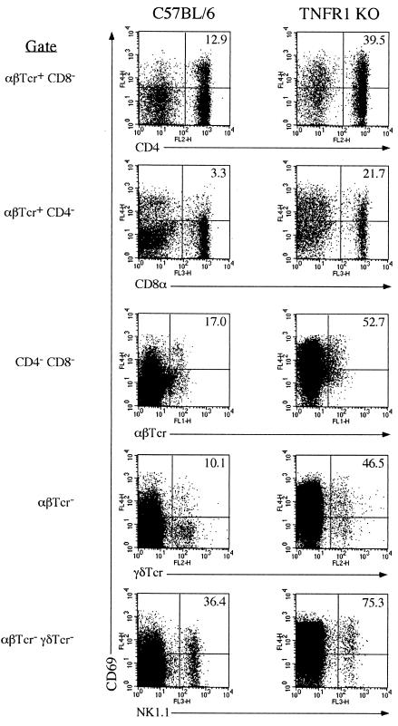
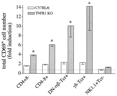
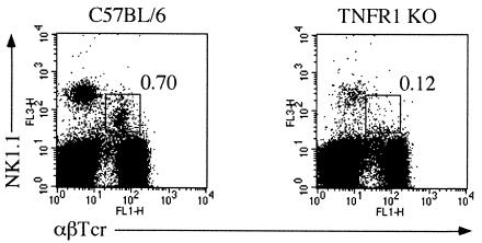
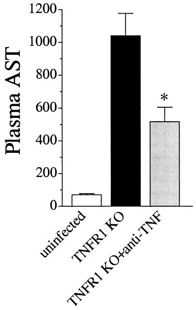
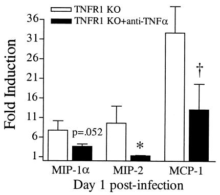
A significant clinical complication of pulmonary infections with Klebsiella pneumoniae is peripheral blood dissemination, resulting in a systemic infection concurrent with the localized pulmonary infection. In this context, little is known about the role of tumor necrosis factor receptor 1 (TNFR1)-mediated innate immune responses during systemic Klebsiella infections. Mice lacking TNFR1 were significantly more susceptible to Klebsiella-induced mortality following intravenous inoculation. Bacterial clearance was impaired in TNFR1-deficient mice at early times following infection. Unexpectedly, bacterial burdens at the onset of mortality (days 2 to 3 postinfection) were not higher in mice lacking TNFR1. However, elevated production of liver-associated proinflammatory cytokines (interleukin-12, tumor necrosis factor alpha [TNF-alpha[, and gamma interferon [IFN-gamma]) and chemokines (MIP-1 alpha, MIP-2, and MCP-1) was observed within the first 24 h of infection. Additionally, excessive plasma-associated IFN-gamma was also observed late in the course of infection (day 3). Spleen cells from day-3 infected TNFR1-deficient mice secreted markedly enhanced levels of IFN-gamma when cultured in vitro. Additionally, there was a marked increase in the total number of activated lymphocyte subsets as indicated by CD69 upregulation. A notable exception was the sharp decrease in the frequency of splenic NK T cells in infected TNFR1 knockout (KO) mice. Anti-TNF-alpha therapy in TNFR1 KO mice significantly reduced chemokine production and liver injury. Combined, these data indicate a dysregulated antibacterial host response following intravenous Klebsiella infection in the absence of TNFR1 signaling, resulting in heightened cytokine production and hyperactivation of specific splenic lymphocyte subsets.


Figures

FIG. 1.
FIG. 1.
Increased mortality in TNFR1- but not TNFR2-deficient mice following intravenous K. pneumoniae infection. (A) TNFR1-deficient mice and their C57BL/6 wild-type controls were intravenously inoculated with 6.8 × 10⁴ K. pneumoniae bacteria, and survival was monitored over 8 days. TNFR1-deficient mice displayed increased mortality as early as day 1 postinfection, and by day 5 all were dead (P < 0.0002 for comparison with control mice). Survival curves were generated from two independent experiments with a total of 18 mice per group. (B) TNFR2-deficient mice and their wild-type controls were intravenously inoculated with 5 × 10⁴ bacteria and monitored for survival over 8 days. Mortality rates were identical for the two groups of animals. Survival curves were generated from three independent experiments with a total of 25 mice per group.
FIG. 2.
FIG. 2.
Liver injury in TNFR1- or TNFR2-deficient mice and wild-type mice following intravenous infection. Plasma AST levels, as an indication of liver cellular injury, were determined 24 h following bacterial infection. All three groups of infected mice displayed a 10-fold increase in AST activity 24 h postinfection. Data are presented as mean AST activities from three independent experiments with a total of 18 to 21 mice per group. Error bars, standard errors of the means.
FIG. 3.
FIG. 3.
Increased production of liver-associated proinflammatory cytokines and chemokines in TNFR1-deficient mice following intravenous Klebsiella infection. Livers from infected TNFR1-deficient mice or infected C57BL/6 mice were removed at 6, 24, 48, and 72 h postinfection, and cytokine or chemokine production was assessed by ELISA as described in Materials and Methods. Induction of cytokines or chemokines is displayed as mean fold induction above that in uninfected controls. Error bars, standard errors of the means. Data were generated from two to four independent experiments with a total of 12 to 24 mice per group. *, P < 0.01; †, P < 0.05.
FIG. 4.
FIG. 4.
Enhanced IFN-γ production by TNFR1-deficient splenocytes following infection. Spleen cells were isolated from uninfected and day-3 infected TNFR1-deficient and C57BL/6 wild-type control animals as described in Materials and Methods. Cells were cultured in vitro with exogenous PMA (50 ng/ml) and ionomycin (100 ng/ml). After 24 h of culture, supernatants were analyzed for IFN-γ secretion. Note that these doses failed to induce detectable IFN-γ secretion from uninfected wild-type or TNFR1-deficient splenocytes. Wild-type infected spleen cells produced IFN-γ at levels slightly above the threshold of detection. In contrast, TNFR1-deficient spleen cells secreted significant levels of IFN-γ following in vitro culture. Data are expressed as mean IFN-γ production from two independent experiments with a total of seven animals, with each individual spleen cultured in duplicate. Error bars, standard errors of the means.
FIG. 5.
FIG. 5.
Increased activation of splenic lymphocyte subsets in TNFR1-deficient mice following infection. Wild-type and TNFR1-deficient mice were intravenously infected, and splenocytes were isolated 3 days postinfection. Cells were stained for CD69 expression on lymphocyte subsets as described in Materials and Methods. Data in dot plots are from events pregated as indicated on the left. The percentage of the cell population of interest (horizontal axis) expressing CD69 is given in the upper right quadrant. Data are representative of two independent experiments with seven mice per group.
FIG. 6.
FIG. 6.
Increased total numbers of activated splenic lymphocyte subsets following infection in TNFR1-deficient mice. Wild-type and TNFR1-deficient mice were intravenously infected, and splenocytes were isolated 3 days postinfection. The percentage of cells expressing CD69 was determined (Fig. 5), and the total number of activated cells was calculated by multiplying the frequency of expression by the total number of splenocytes of that subset. Infected TNFR1-deficient spleens contained significantly more activated DN-αβ T cells and γδ T cells and, to a lesser extent, more CD4+ T cells and CD8+ T cells than infected wild-type spleens (*, P < 0.01). Dotted line indicates baseline number of cells prior to infection. Data are representative of two independent experiments with seven mice per group.
FIG. 7.
FIG. 7.
Decreased numbers of splenic NK T cells in TNFR1-deficient mice following infection. The frequency of splenic NK T cells (defined as NK1.1+ αβTcr+ γδTcr) was determined as described in Materials and Methods. The frequencies of splenic NK T cells in infected wild-type and TNFR1-deficient mice are given within the appropriate dot plots. Data are representative of two independent experiments with seven mice per group.
FIG. 8.
FIG. 8.
Reduced liver injury in TNFR1-deficient mice following TNF-α neutralization. An anti-TNF-α monoclonal antibody was intraperitoneally administered 2 h prior to infection. TNFR1 knockout (KO) mice were then intravenously inoculated with K. pneumoniae and analyzed 24 h postinfection. Plasma AST levels, as an indication of liver cellular injury, were determined 24 h following bacterial infection. Anti-TNF-α treatment reduced AST levels by 50% in TNFR1-deficient mice following infection (*, P < 0.01).
FIG. 9.
FIG. 9.
Decreased chemokine production in TNFR1-deficient mice following TNF-α neutralization. TNFR1-deficient mice were pretreated with anti-TNF-α 2 h prior to intravenous K. pneumoniae infection. Livers were excised 24 h postinfection, and induction of the chemokines MIP-1α, MIP-2, and MCP-1 was determined by ELISA as described in Materials and Methods. Anti-TNF-α treatment significantly reduced the excessive induction of MIP-2 (*, P < 0.001) and MCP-1 (†, P < 0.05), along with a consistent trend for reduced MIP-1α levels (P = 0.052), in TNFR1-deficient mice. Data are presented as mean fold induction (over levels in uninfected animals) from three independent experiments with a total of 15 mice per group. Error bars, standard errors of the means.

Similar articles

Cited by

References

    1. Allendoerfer, R., and G. S. Deepe, Jr. 2000. Regulation of infection with Histoplasma capsulatum by TNFR1 and -2. J. Immunol. 165:2657-2664. - PubMed
    1. Bank, U., and S. Ansorge. 2001. More than destructive: neutrophil-derived serine proteases in cytokine bioactivity control. J. Leukoc. Biol. 69:197-206. - PubMed
    1. Burwen, D. R., S. N. Banerjee, and R. P. Gaynes. 1994. Ceftazidime resistance among selected nosocomial gram-negative bacilli in the United States. National Nosocomial Infections Surveillance System. J. Infect. Dis. 170:1622-1625. - PubMed
    1. Carnaud, C., D. Lee, O. Donnars, S. H. Park, A. Beavis, Y. Koezuka, and A. Bendelac. 1999. Cross-talk between cells of the innate immune system: NKT cells rapidly activate NK cells. J. Immunol. 163:4647-4650. - PubMed
    1. Chosay, J. G., N. A. Essani, C. J. Dunn, and H. Jaeschke. 1997. Neutrophil margination and extravasation in sinusoids and venules of liver during endotoxin-induced injury. Am. J. Physiol. 272:G1195-G1200. - PubMed

Publication types

MeSH terms