. 2003 Sep;71(9):5296-305.
doi: 10.1128/IAI.71.9.5296-5305.2003.

Characterization of Pseudomonas aeruginosa exoenzyme S as a bifunctional enzyme in J774A.1 macrophages

Affiliations

Characterization of Pseudomonas aeruginosa exoenzyme S as a bifunctional enzyme in J774A.1 macrophages

Claudia L Rocha et al. Infect Immun. 2003 Sep.

Abstract

Pseudomonas aeruginosa exoenzyme S (ExoS) is a type III secretion (TTS) effector, which includes both a GTPase-activating protein (GAP) activity toward the Rho family of low-molecular-weight G (LMWG) proteins and an ADP-ribosyltransferase (ADPRT) activity that targets LMWG proteins in the Ras, Rab, and Rho families. The coordinate function of both activities of ExoS in J774A.1 macrophages was assessed by using P. aeruginosa strains expressing and translocating wild-type ExoS or ExoS defective in GAP and/or ADPRT activity. Distinct and coordinated functions were identified for both domains. The GAP activity was required for the antiphagocytic effect of ExoS and was linked to interference of lamellopodium and membrane ruffle formation. Alternatively, the ADPRT activity of ExoS altered cellular adherence and morphology and was linked to effects on filopodium formation. The cellular mechanism of ExoS GAP activity included an inactivation of Rac1 function, as determined in p21-activated kinase 1-glutathione S-transferase (GST) pull-down assays. The ADPRT activity of ExoS targeted Ras and RalA but not Rab or Rho proteins, and Ral binding protein 1-GST pull-down assays identified an effect of ExoS ADPRT activity on RalA activation. The results from these studies confirm the bifunctional nature of ExoS activity within macrophages when translocated by TTS.


Figures

FIG. 1.
FIG. 1.
Cytotoxic effects of ExoS GAP and ADPRT activities on the J774A.1 cell line. J774A.1 cells were seeded at 105 cells/ml and cultured for 72 h, to 70% confluency, prior to coculture for 2, 4, and 6 h, with no bacteria (0) or with 107 CFU of strain PA103ΔUT expressing the indicated ExoS construct (described in Table 1) per ml. (A) Effect on morphology. After 5 h of coculture, bacteria were removed, cells were washed once with PBS, and medium containing antibiotic was added to limit bacterial growth. Cells were visualized by phase-contrast microscopy. Cell rounding was more evident in cells cocultured with bacteria expressing ADPRT-active ExoS (ExoS and R146A). Magnification, ×32. (B) Effect on adherence. At 6 h, when loss of cell adherence occurred, nonadherent and attached cells were recovered, washed with PBS and quantified by using a trypan blue stain. Results are expressed as the percentage of total cells that are nonadherent. The means and standard errors from five independent experiments are shown, and an asterisk indicates a statistically significant difference in adherence (P = 0.0002 for ExoS and P = 0.0001 for R146A relative to control cells not treated with bacteria, P = 0.01 for ExoS and P = 0.003 for R146A relative to the pUCP vector control, and P = 0.0001 for ExoS and R146A relative to E379A/E381A and P = 0.02 for ExoS and R146A relative to R146A/E379A/E381A).
FIG. 2.
FIG. 2.
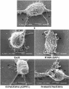
Analysis of J774A.1 cell morphology by SEM. J774A.1 cells were grown on Thermanox coverslips and cocultured for 2.5 h with the indicated ExoS-expressing PA103ΔUT strain. Cells were fixed with 2% cacodylate glutaraldehyde, followed by 2% osmium tetroxide. Samples were rinsed, dehydrated with ethanol, and then incubated with hexamethyldisilazane until dry, mounted, sputter coated with gold palladium, and examined with a JEOL 5410 scanning electron microscope. Representative pictures of the predominant phenotype associated with each mutant are shown. Control cells (0) showed normal macrophage morphology. Cells cocultured with PA103ΔUT expressing ExoS showed decreased lamellipodia and membrane ruffling (70.5%; 12 of 17 cells) and decreased filopodia (58.8%; 10 of 17 cells). Cells cocultured with the R146A-GAP mutant showed enhanced lamellipodia and ruffles (100%; 10 of 10 cells) and no filopodia (70%; 7 of 10 cells). Cells cocultured with the E379A/E381A-ADPRT mutant showed no lamellipodia and ruffles (60%; 6 of 10 cells) and restored filopodia (70%; 7 of 10 cells). Cells cocultured with the R146A/E379A/E381A-GAP/ADPRT mutant showed restored lamellipodia, membrane ruffles, and filopodia (100%; nine of nine cells). Arrowheads identify lamellipodia, and arrows identify filopodia. Bar, 5 μm.
FIG. 3.
FIG. 3.
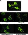
Antiphagocytic activity of ExoS determined by a double-immunofluorescence assay. (A) Fluorescence microscopy. J774A.1 cells were seeded on slides, cultured as described for Fig. 1, and then cocultured for 2.5 h with no bacteria (0) or with 107 CFU of the indicated PA103ΔUT strain per ml. Cells were washed, fixed with 3% paraformaldehyde, and blocked. Extracellular bacteria were stained first with guinea pig anti-P. aeruginosa IgG polyclonal antibody and visualized with a goat anti-guinea pig IgG-SP-biotin conjugate, followed by Extravidin-FITC conjugate (green bacteria). Intracellular bacteria were stained second by permeabilizing cells with 100% methanol at −20°C and staining as described above, with the exception that Extravidin-tetramethyl rhodamine isocyanate was used (red bacteria). Slides were mounted and examined by fluorescence microscopy for cells with associated extracellular bacteria (green) and/or intracellular bacteria (red). Magnification, ×100. (B) Quantification of intracellular bacteria. The experimental results from panel A were quantified as the percentage of cells with internalized bacteria, with the means and standard errors of analyses performed in triplicate in three independent experiments represented. An asterisk indicates a statistically significant difference for the R146A-GAP and R146A/E379A/E381A-GAP/ADPRT mutants relative to control cells (0), as well as to all of the other strains analyzed (P ≤ 0.05).
FIG. 3.
FIG. 3.
Antiphagocytic activity of ExoS determined by a double-immunofluorescence assay. (A) Fluorescence microscopy. J774A.1 cells were seeded on slides, cultured as described for Fig. 1, and then cocultured for 2.5 h with no bacteria (0) or with 107 CFU of the indicated PA103ΔUT strain per ml. Cells were washed, fixed with 3% paraformaldehyde, and blocked. Extracellular bacteria were stained first with guinea pig anti-P. aeruginosa IgG polyclonal antibody and visualized with a goat anti-guinea pig IgG-SP-biotin conjugate, followed by Extravidin-FITC conjugate (green bacteria). Intracellular bacteria were stained second by permeabilizing cells with 100% methanol at −20°C and staining as described above, with the exception that Extravidin-tetramethyl rhodamine isocyanate was used (red bacteria). Slides were mounted and examined by fluorescence microscopy for cells with associated extracellular bacteria (green) and/or intracellular bacteria (red). Magnification, ×100. (B) Quantification of intracellular bacteria. The experimental results from panel A were quantified as the percentage of cells with internalized bacteria, with the means and standard errors of analyses performed in triplicate in three independent experiments represented. An asterisk indicates a statistically significant difference for the R146A-GAP and R146A/E379A/E381A-GAP/ADPRT mutants relative to control cells (0), as well as to all of the other strains analyzed (P ≤ 0.05).
FIG. 4.
FIG. 4.
Substrate modification by ExoS ADPRT activity in J774A.1 cells. J774A.1 cells were seeded and cocultured for 5 h with the indicated ExoS-expressing PA103ΔUT strain, as described for Fig. 1. For the analysis of Ras modification, cells were lysed in TBS-TDS buffer (10 mM Tris [pH 7.4], 140 mM NaCl, 1% Triton X 100, 0.5% sodium deoxycholate, 0.1% SDS) for 30 min on ice. Lysates were cleared by centrifugation, and Ras was immunoprecipitated with monoclonal Y13-259 Ras antibody. For analyses of all the other LMWG proteins, cells were scraped, washed with PBS and lysed as described in Materials and Methods. Samples were resolved by SDS-PAGE, transferred to polyvinylidene difluoride membranes, and probed with antibodies specific for the indicated LMWG protein. Blots were visualized by enhanced chemiluminescence. The mobilities of modified (M) and unmodified (U) proteins are indicated. The results are representative of those from analyses performed in multiple independent studies. TTS-translocated ExoS substrate modification of J774A.1 cells is compared with the more extensive substrate modification previously reported for HT-29 human epithelial cells (8).
FIG. 5.
FIG. 5.
Analysis of Rac1 and RalA activation. J774A.1 cells were grown and cocultured with the indicated PA103ΔUT strains for 5 h, as for Fig. 1. Bacteria were removed and the cells treated as follows. (A) To detect GTP-active Rac1, cells were lysed and incubated with PAK-GST bound to GSH beads. Proteins bound to beads were resolved by SDS-PAGE, and Rac1 was detected by immunoblot analyses with a Rac1-specific antibody. (B) To detect GTP-active RalA, cells were lysed in RBD-lysis buffer and incubated with the RBD-GST bound to GSH beads. RalA binding to beads was examined as described above but detected with a RalA-specific antibody. The results are representative of those from three independent studies. The ratio of active Rac1-GTP or RalA-GTP to total Rac1 or RalA, respectively, in each lysate was quantified based on densitometry analysis and normalized relative to untreated control cells. The mean (standard deviation) for each condition is represented. Ratios of greater than one, relative to untreated control cells, observed in response to some bacterial strains are predicted to reflect the activation of the LMWG proteins in J774A.1 cells upon exposure to bacteria.

Similar articles

Cited by

References

    1. Aktories, K., C. Mohr, and G. Koch. 1992. Clostridium botulinum C3 ADP-ribosyltransferase. Curr. Top. Microbiol. Immunol. 175:115-131. - PubMed
    1. Allen, W. E., G. E. Jones, J. W. Pollard, and A. J. Ridley. 1997. Rho, Rac and Cdc42 regulate actin organization and cell adhesion in macrophages. J. Cell Sci. 110:707-720. - PubMed
    1. Caron, E., and A. Hall. 1998. Identification of two distinct mechanisms of phagocytosis controlled by different Rho GTPases. Science 282:1717-1721. - PubMed
    1. Coburn, J., and D. W. Frank. 1999. Macrophages and epithelial cells respond differently to the Pseudomonas aeruginosa type III secretion system. Infect. Immun. 67:3151-3154. - PMC - PubMed
    1. Coburn, J., R. T. Wyatt, B. H. Iglewski, and D. M. Gill. 1989. Several GTP-binding proteins, including p21c-H-ras, are preferred substrates of Pseudomonas aeruginosa exoenzyme S. J. Biol. Chem. 264:9004-9008. - PubMed

Publication types

MeSH terms