. 2003 Sep;47(9):2892-6.
doi: 10.1128/AAC.47.9.2892-2896.2003.

Resistance to the peptidyl transferase inhibitor tiamulin caused by mutation of ribosomal protein l3

Affiliations

Resistance to the peptidyl transferase inhibitor tiamulin caused by mutation of ribosomal protein l3

Jacob Bøsling et al. Antimicrob Agents Chemother. 2003 Sep.

Abstract

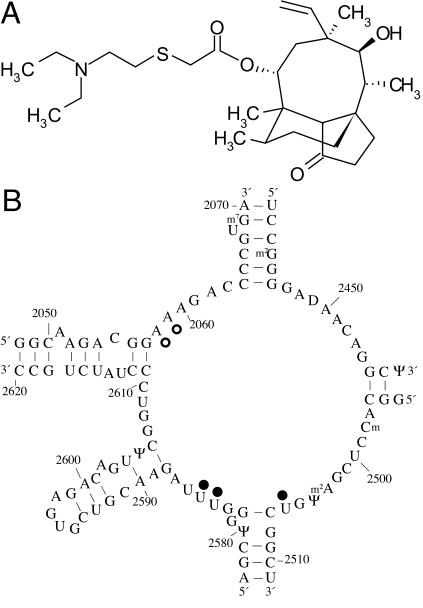
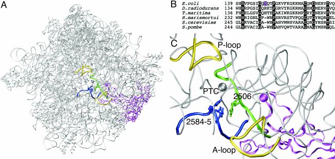
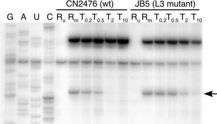
The antibiotic tiamulin targets the 50S subunit of the bacterial ribosome and interacts at the peptidyl transferase center. Tiamulin-resistant Escherichia coli mutants were isolated in order to elucidate mechanisms of resistance to the drug. No mutations in the rRNA were selected as resistance determinants using a strain expressing only a plasmid-encoded rRNA operon. Selection in a strain with all seven chromosomal rRNA operons yielded a mutant with an A445G mutation in the gene coding for ribosomal protein L3, resulting in an Asn149Asp alteration. Complementation experiments and sequencing of transductants demonstrate that the mutation is responsible for the resistance phenotype. Chemical footprinting experiments show a reduced binding of tiamulin to mutant ribosomes. It is inferred that the L3 mutation, which points into the peptidyl transferase cleft, causes tiamulin resistance by alteration of the drug-binding site. This is the first report of a mechanism of resistance to tiamulin unveiled in molecular detail.


Figures

FIG. 1.
FIG. 1.
(A) Chemical structure of tiamulin; (B) chemical footprint of tiamulin on 23S rRNA indicated on the secondary structure of domain V of E. coli 23S rRNA. Nucleotides with altered reactivities in the presence of tiamulin are indicated with filled circles (protections) or open circles (enhancements).
FIG. 2.
FIG. 2.
(A) The position of ribosomal protein L3 in the large ribosomal subunit. The subunit is shown in the crown view, where the peptidyl transferase center is in the middle of the figure. Ribosomal protein L3 is shown as a magenta ribbon, and the position of the mutation is marked with a sphere of the same color. The other highlighted items and their coloring are as described for panel C. Other ribosomal proteins and rRNA in the large ribosomal subunit are depicted as gray tubes. The coordinates of the H. marismortui 50S subunit are used (PDB accession number 1JJ2). (B) Sequence comparison of various L3 ribosomal proteins in the regions flanking the E. coli Asn149Asp mutation. The other L3 sequences are from the bacteria D. radiodurans and Thermotoga maritima, the archaeon H. marismortui, and the eukaryotes S. cerevisiae and Schizosaccharomyces pombe. The position of the mutation in E. coli is highlighted in magenta, and positions of amino acid identity are highlighted in black. (C) Close-up view of the location of the L3 mutation relative to rRNA nucleotides at the peptidyl transferase center. Ribosomal protein L3 is depicted as in panel A. The nucleotides in the tiamulin footprint are shown in green (U2506) and blue (U2584 and U2585), and the respective regions of 23S rRNA flanking these nucleotides are highlighted in the same color (nucleotides are labeled using E. coli numbering). The peptidyl transferase center (PTC) is indicated by a gray sphere centered on the N-3 atom of A2486 (A2451 in E. coli numbering). The A and P loops of 23S rRNA, bound transiently by tRNAs in the A and P sites, respectively, are depicted as yellow tubes. The gray tubes represent parts of domain V of H. marismortui 23S rRNA.
FIG. 3.
FIG. 3.
Gel autoradiogram showing the protection effect at nucleotide U2506 in E. coli 23S rRNA caused by tiamulin binding to CN2476 (wild-type [wt]) and JB5 (L3 mutant) ribosomes. Dideoxy sequencing lanes are indicated by G, A, U, and C. Lanes are labeled to denote reactions with chemically unmodified 70S ribosomes in the absence of tiamulin (Ru), 70S ribosomes modified with CMCT in the absence of tiamulin (Rm), or 70S ribosomes modified in the presence of tiamulin (T0.2, T0.5, T2 or T10, where the subscript indicates the drug concentration [micromolar]). The arrow shows the altered reactivity at position U2506.

References

    1. Asai, T., D. Zaporojets, C. Squires, and C. L. Squires. 1999. An Escherichia coli strain with all chromosomal rRNA operons inactivated: complete exchange of rRNA genes between bacteria. Proc. Natl. Acad. Sci. USA 96:1971-1976. - PMC - PubMed
    1. Ban, N., P. Nissen, J. Hansen, P. Moore, and T. A. Steitz. 2000. The complete atomic structure of the large ribosomal subunit at 2.4 Å resolution. Science 289:905-920. - PubMed
    1. Böck, A., F. Turnowsky, and G. Högenauer. 1982. Tiamulin resistance mutations in Escherichia coli. J. Bacteriol. 151:1253-1260. - PMC - PubMed
    1. Douthwaite, S. R., T. Powers, J. Y. Lee, and H. F. Noller. 1989. Defining the structural requirements for a helix in 23 S ribosomal RNA that confers erythromycin resistance. J. Mol. Biol. 209:655-665. - PubMed
    1. Fried, H. M., and J. R. Warner. 1981. Cloning of yeast gene for trichodermin resistance and ribosomal protein L3. Proc. Natl. Acad. Sci. USA 78:238-242. - PMC - PubMed

Publication types

LinkOut - more resources