

The aminoglycoside 6'-N-acetyltransferase type Ib encoded by Tn1331 is evenly distributed within the cell's cytoplasm
- PMID: 12936992
- PMCID: PMC182613
- DOI: 10.1128/AAC.47.9.2897-2902.2003
The aminoglycoside 6'-N-acetyltransferase type Ib encoded by Tn1331 is evenly distributed within the cell's cytoplasm
Abstract
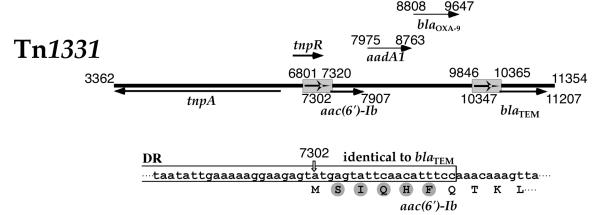
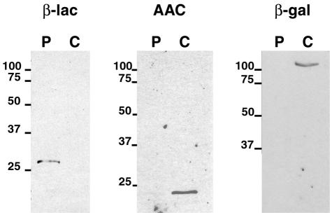
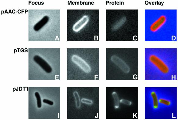
The multiresistance transposon Tn1331, which mediates resistance to several aminoglycosides and beta-lactams, includes the aac(6')-Ib, aadA1, bla(OXA-9), and bla(TEM-1) genes. The nucleotide sequence of aac(6')-Ib includes a region identical to that of the bla(TEM-1) gene. This region encompasses the promoter and the initiation codon followed by 15 nucleotides. Since there were three possible translation initiation sites, the amino acid sequence at the N terminus of the aminoglycoside 6'-N-acetyltransferase type Ib [AAC(6')-Ib] was determined and was found to be SIQHF. This result indicated that aac(6')-Ib includes a translational fusion: the first five amino acids of the leader peptide of the TEM beta-lactamase are fused to the rest of the AAC(6')-Ib protein. This gene fusion could have formed during the genesis of Tn1331 as a consequence of the generation of a 520-nucleotide duplication (M. E. Tolmasky, Plasmid 24:218-226, 1990). An identical gene isolated from a Serratia marcescens strain has been previously described (G. Tran van Nhieu and E. Collatz, J. Bacteriol. 169:5708-5714, 1987). Extraction of the periplasmic proteins of E. coli harboring aac(6')-Ib by spheroplast formation showed that most of the AAC(6')-Ib protein is present in the cytoplasm. A genetic fusion to phoA confirmed these results. AAC(6')-Ib was shown to be evenly distributed inside the cell's cytoplasm by fluorescent microscopy with an AAC(6')-Ib-cyan fluorescent protein fusion.
Figures
References
-
- Bachmann, B. 1996. Derivations and genotypes of some mutant derivatives of Escherichia coli K-12, p. 2460-2495. In F. C. Neidhardt, R. Curtiss III, J. L. Ingraham, E. C. C. Lin, K. B. Low, B. Magasanik, W. S. Reznikoff, M. Riley, M. Schaechter, and H. E. Umbarger (ed.), Escherichia coli and Salmonella: cellular and molecular biology, 2nd ed., vol. 2. ASM Press, Washington, D.C.
-
- Bowden, G. A., and G. Georgiou. 1990. Folding and aggregation of beta-lactamase in the periplasmic space of Escherichia coli. J. Biol. Chem. 265:16760-16766. - PubMed
-
- Boyer, H. W., and D. Roulland-Dussoix. 1969. A complementation analysis of the restriction and modification of DNA in Escherichia coli. J. Mol. Biol. 41:459-472. - PubMed
Publication types
MeSH terms
Substances
Associated data
- Actions
Grants and funding
LinkOut - more resources
Full Text Sources
