

Retinal angiogenesis is mediated by an interaction between the angiotensin type 2 receptor, VEGF, and angiopoietin
- PMID: 12937129
- PMCID: PMC1868261
- DOI: 10.1016/S0002-9440(10)63448-7
Retinal angiogenesis is mediated by an interaction between the angiotensin type 2 receptor, VEGF, and angiopoietin
Abstract
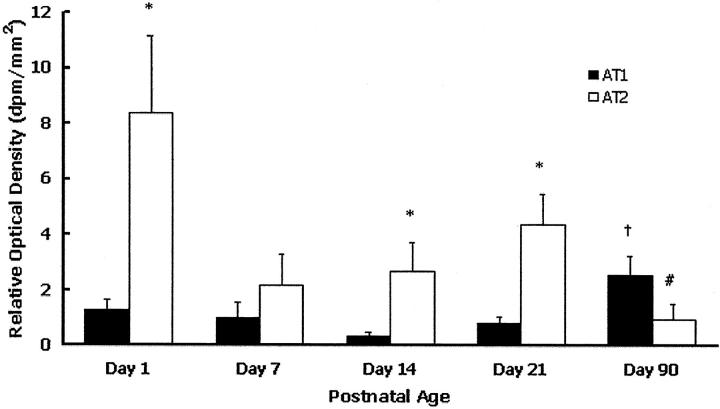
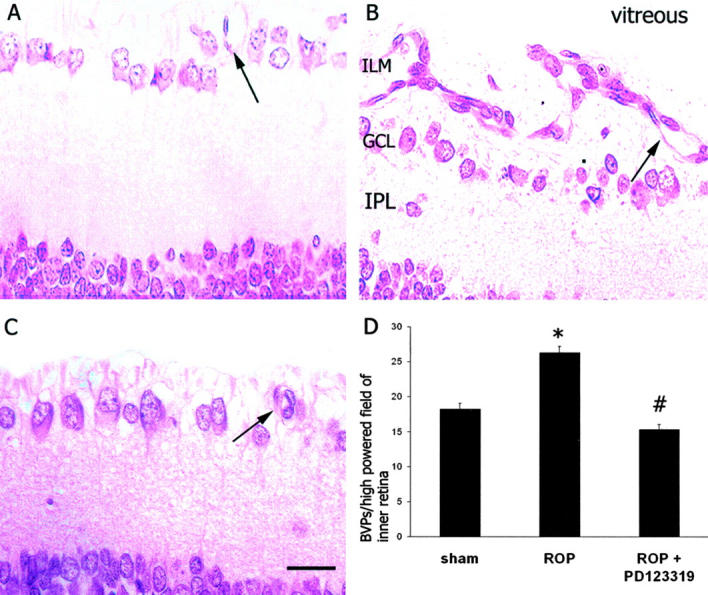
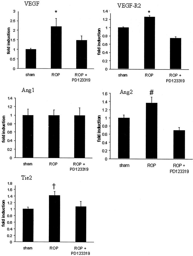
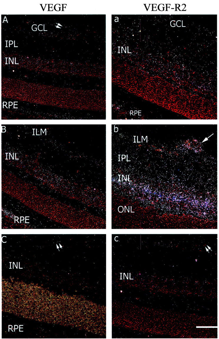
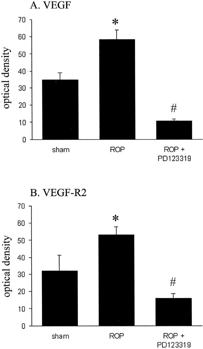
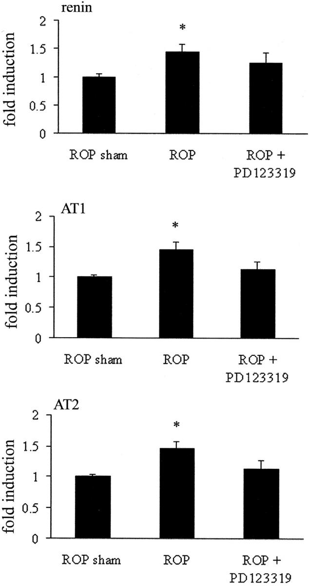
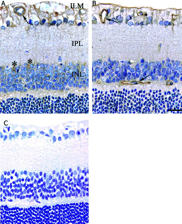
There is evidence that angiotensin II, vascular endothelial growth factor (VEGF), angiopoietins, and their cognate receptors participate in retinal angiogenesis. We investigated whether angiotensin type 2-receptor blockade (AT2-RB) reduces retinal angiogenesis and alters the expression of VEGF/VEGF-R2 and angiopoietin-Tie2. Retinopathy of prematurity (ROP) was induced in Sprague Dawley (SD) rats by exposure to 80% oxygen from postnatal (P) days 0 to 11, followed by 7 days in room air. ROP shams were in room air from P0-18. A group of ROP rats received the AT2-RB, PD123319, by mini-osmotic pump (5 mg/kg/day) from P11-18 (angiogenesis period). Evaluation of the retinal status of the AT2 receptor indicated that this receptor, as assessed by real-time PCR, immunohistochemistry, and in vitro autoradiography, was present in the retina, was more abundant than the AT1 receptor in the neonatal retina, and was increased in the ROP model. AT2-RB reduced retinal angiogenesis. VEGF and VEGF-R2 mRNA were increased in ROP and localized to blood vessels, ganglion cells, and the inner nuclear layer, and were decreased by PD123319. Angiopoietin2 and Tie2, but not angiopoietin1 mRNA were increased with ROP, and angiopoietin2 was reduced with PD123319. This study has identified a potential retinoprotective role for AT2-RB possibly mediated via interactions with VEGF- and angiopoietin-dependent pathways.
Figures
Similar articles
-
Colocalization of Tie2, angiopoietin 2 and vascular endothelial growth factor in fibrovascular membrane from patients with retinopathy of prematurity.Ophthalmic Res. 2003 Jul-Aug;35(4):217-23. doi: 10.1159/000071173. Ophthalmic Res. 2003. PMID: 12815197
-
Vascular endothelial growth factor and its receptors in control and diabetic rat eyes.Lab Invest. 1998 Aug;78(8):1017-27. Lab Invest. 1998. PMID: 9714188
-
Hypoxia inducible factor-1alpha is increased in ischemic retina: temporal and spatial correlation with VEGF expression.Invest Ophthalmol Vis Sci. 1999 Jan;40(1):182-9. Invest Ophthalmol Vis Sci. 1999. PMID: 9888442
-
Studies on the pathogenesis of avascular retina and neovascularization into the vitreous in peripheral severe retinopathy of prematurity (an american ophthalmological society thesis).Trans Am Ophthalmol Soc. 2010 Dec;108:96-119. Trans Am Ophthalmol Soc. 2010. PMID: 21212851 Free PMC article. Review.
-
Vascular endothelial growth factor gene regulation and action in diabetic retinopathy.Ophthalmol Clin North Am. 2002 Mar;15(1):69-79. doi: 10.1016/s0896-1549(01)00010-4. Ophthalmol Clin North Am. 2002. PMID: 12064083 Review.
Cited by
-
Renin-Angiotensin system hyperactivation can induce inflammation and retinal neural dysfunction.Int J Inflam. 2012;2012:581695. doi: 10.1155/2012/581695. Epub 2012 Mar 22. Int J Inflam. 2012. PMID: 22536545 Free PMC article.
-
Safety and Efficacy of Systemic Anti-Scg3 Therapy to Treat Oxygen-Induced Retinopathy.Front Biosci (Landmark Ed). 2022 Apr 19;27(4):130. doi: 10.31083/j.fbl2704130. Front Biosci (Landmark Ed). 2022. PMID: 35468689 Free PMC article.
-
The significance of neuronal and glial cell changes in the rat retina during oxygen-induced retinopathy.Doc Ophthalmol. 2010 Feb;120(1):67-86. doi: 10.1007/s10633-009-9193-6. Epub 2009 Sep 8. Doc Ophthalmol. 2010. PMID: 19763649 Review.
-
Short hairpin RNA-mediated knockdown of VEGFA in Müller cells reduces intravitreal neovascularization in a rat model of retinopathy of prematurity.Am J Pathol. 2013 Sep;183(3):964-74. doi: 10.1016/j.ajpath.2013.05.011. Am J Pathol. 2013. PMID: 23972394 Free PMC article.
-
Regulation of angiotensin II receptors and extracellular matrix turnover in human retinal pigment epithelium: role of angiotensin II.Am J Physiol Cell Physiol. 2008 Dec;295(6):C1633-46. doi: 10.1152/ajpcell.00092.2008. Epub 2008 Oct 15. Am J Physiol Cell Physiol. 2008. PMID: 18923060 Free PMC article.
References
-
- Klein R, Klein B, Moss S: Epidemiology of proliferative diabetic retinopathy. Diabetes Care 1992, 15:1875-1891 - PubMed
-
- Fryczkowski A, Hodes B, Walker J: Diabetic choroidal and iris vasculature scanning electron microscopy findings. Int Ophthalmol Clin 1989, 13:269-279 - PubMed
-
- Phelps DL: Retinopathy of prematurity. Pediatr Rev 1995, 16:50-56 - PubMed
-
- Schalekamp MADH: Renin-angiotensin system components and endothelial proteins as markers of diabetic microvascular disease. Clin Invest 1993, 71:S3-S6 - PubMed
-
- Danser AHJ, Van den Dorpel MA, Deinum J, Derkx FHM, Franken AAM, Peperkamp E, de Jong PTVM, Schalekamp MADH: Renin, prorenin, and immunoreactive renin in vitreous fluid from eyes with and without diabetic retinopathy. J Clin Endocrinol Metab 1989, 68:160-167 - PubMed
Publication types
MeSH terms
Substances
LinkOut - more resources
Full Text Sources
Other Literature Sources
Medical
Research Materials
Miscellaneous
