

Vascular smooth muscle cells orchestrate the assembly of type I collagen via alpha2beta1 integrin, RhoA, and fibronectin polymerization
- PMID: 12937145
- PMCID: PMC1868248
- DOI: 10.1016/s0002-9440(10)63464-5
Vascular smooth muscle cells orchestrate the assembly of type I collagen via alpha2beta1 integrin, RhoA, and fibronectin polymerization
Abstract
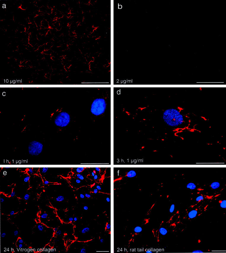
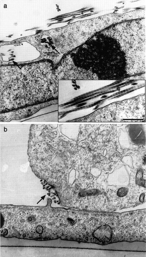
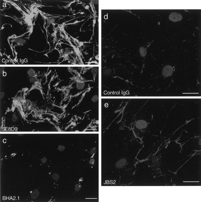
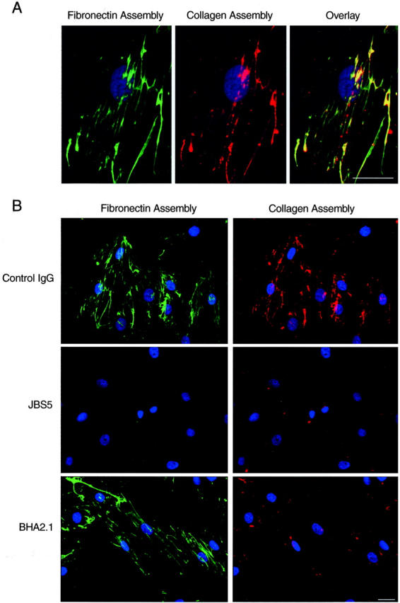
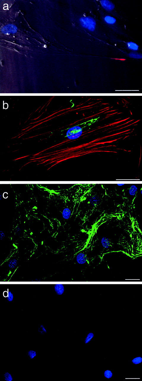
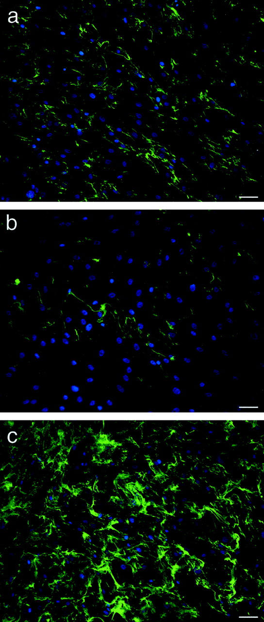
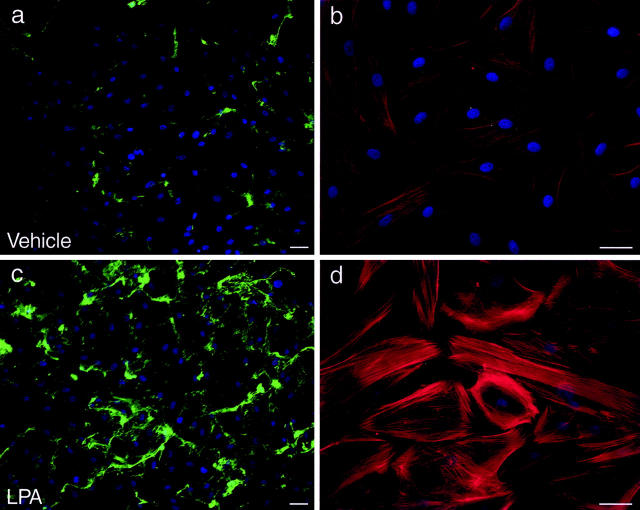
Assembly of collagen into fibrils is widely studied as a spontaneous and entropy-driven process. To determine whether vascular smooth muscle cells (SMCs) impact the formation of collagen fibrils, we microscopically tracked the conversion of soluble to insoluble collagen in human SMC cultures, using fluorescent type I collagen at concentrations less than that which supported self-assembly. Collagen microaggregates were found to form on the cell surface, initially as punctate collections and then as an increasingly intricate network of fibrils. These fibrils displayed 67-nm periodicity and were found in membrane-delimited cellular invaginations. Fibril assembly was inhibited by an anti-alpha2beta1 integrin antibody and accelerated by an alpha2beta1 integrin antibody that stimulates a high-affinity binding state. Newly assembled collagen fibrils were also found to co-localize with newly assembled fibronectin fibrils. Moreover, inhibition of fibronectin assembly with an anti-alpha5beta1 integrin antibody completely inhibited collagen assembly. Collagen fibril formation was also linked to the cytoskeleton. Fibrils formed on the stretched tails of SMCs, ran parallel to actin microfilament bundles, and formed poorly on SMCs transduced with retrovirus containing cDNA for dominant-negative RhoA and robustly on SMCs expressing constitutively active RhoA. Lysophosphatidic acid, which activates RhoA and stimulates fibronectin assembly, stimulated collagen fibril formation, establishing for the first time that collagen polymerization can be regulated by soluble agonists of cell function. Thus, collagen fibril formation is under close cellular control and is dynamically integrated with fibronectin assembly, opening new possibilities for modifying collagen deposition.
Figures
Similar articles
-
Lipid incorporation inhibits Src-dependent assembly of fibronectin and type I collagen by vascular smooth muscle cells.Circ Res. 2009 Apr 10;104(7):832-41. doi: 10.1161/CIRCRESAHA.108.187302. Epub 2009 Feb 19. Circ Res. 2009. PMID: 19229059
-
alpha5beta1 integrin expression and luminal edge fibronectin matrix assembly by smooth muscle cells after arterial injury.Am J Pathol. 2000 Feb;156(2):453-65. doi: 10.1016/s0002-9440(10)64750-5. Am J Pathol. 2000. PMID: 10666375 Free PMC article.
-
Fibroblast growth factor-2 potentiates vascular smooth muscle cell migration to platelet-derived growth factor: upregulation of alpha2beta1 integrin and disassembly of actin filaments.Circ Res. 1997 May;80(5):627-37. doi: 10.1161/01.res.80.5.627. Circ Res. 1997. PMID: 9130443
-
Collagen Assembly at the Cell Surface: Dogmas Revisited.Cells. 2021 Mar 16;10(3):662. doi: 10.3390/cells10030662. Cells. 2021. PMID: 33809734 Free PMC article. Review.
-
Collagen fibrillogenesis: fibronectin, integrins, and minor collagens as organizers and nucleators.Curr Opin Cell Biol. 2008 Oct;20(5):495-501. doi: 10.1016/j.ceb.2008.06.008. Epub 2008 Jul 30. Curr Opin Cell Biol. 2008. PMID: 18640274 Free PMC article. Review.
Cited by
-
Impact of type-1 collagen hydrogel density on integrin-linked morphogenic response of SH-SY5Y neuronal cells.RSC Adv. 2021 Oct 7;11(52):33124-33135. doi: 10.1039/d1ra05257h. eCollection 2021 Oct 4. RSC Adv. 2021. PMID: 35493559 Free PMC article.
-
Fibrillin assembly requires fibronectin.Mol Biol Cell. 2009 Feb;20(3):846-58. doi: 10.1091/mbc.e08-08-0830. Epub 2008 Nov 26. Mol Biol Cell. 2009. PMID: 19037100 Free PMC article.
-
The Role of Integrin Receptor's α and β Subunits of Mouse Mesenchymal Stem Cells on the Interaction of Marine-Derived Blacktip Reef Shark (Carcharhinus melanopterus) Skin Collagen.Int J Mol Sci. 2023 May 23;24(11):9110. doi: 10.3390/ijms24119110. Int J Mol Sci. 2023. PMID: 37298062 Free PMC article.
-
Biomechanics of soft biological tissues and organs, mechanobiology, homeostasis and modelling.J R Soc Interface. 2025 Jan;22(222):20240361. doi: 10.1098/rsif.2024.0361. Epub 2025 Jan 29. J R Soc Interface. 2025. PMID: 39876788 Free PMC article. Review.
-
Isotropic imaging across spatial scales with axially swept light-sheet microscopy.Nat Protoc. 2022 Sep;17(9):2025-2053. doi: 10.1038/s41596-022-00706-6. Epub 2022 Jul 13. Nat Protoc. 2022. PMID: 35831614 Free PMC article. Review.
References
-
- Jaenisch R, Harbers K, Schnieke A, Lohler J, Chumakov I, Jahner D, Grotkopp D, Hoffmann E: Germline integration of Moloney murine leukemia virus at the Mov13 locus leads to recessive lethal mutation and early embryonic death. Cell 1983, 32:209-216 - PubMed
-
- Breindl M, Harbers K, Jaenisch R: Retrovirus-induced lethal mutation in collagen I gene of mice is associated with an altered chromatin structure. Cell 1984, 38:9-16 - PubMed
-
- Trelstad RL, Silver RH: Matrix assembly. HED eds. Cell Biology of the Extracellular Matrix. 1981:pp 179-216 Plenum Publishing Corp. New York
-
- Gross J, Kirk D: Heat precipitation of collagen from neutral salt solutions: some rate regulating factors. J Biol Chem 1958, 233:355-360 - PubMed
Publication types
MeSH terms
Substances
LinkOut - more resources
Full Text Sources
Other Literature Sources
Miscellaneous
