. 2003 Sep;163(3):1069-80.
doi: 10.1016/S0002-9440(10)63466-9.

Complement C5 in experimental autoimmune encephalomyelitis (EAE) facilitates remyelination and prevents gliosis

Affiliations

Complement C5 in experimental autoimmune encephalomyelitis (EAE) facilitates remyelination and prevents gliosis

Susanna H Weerth et al. Am J Pathol. 2003 Sep.

Abstract

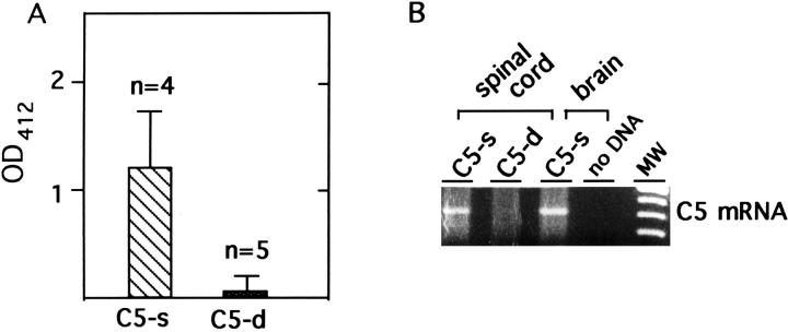
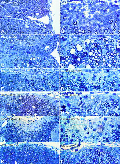
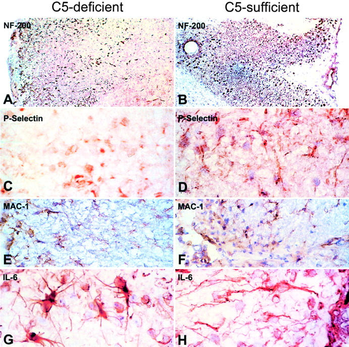
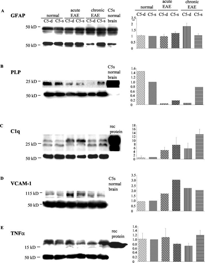
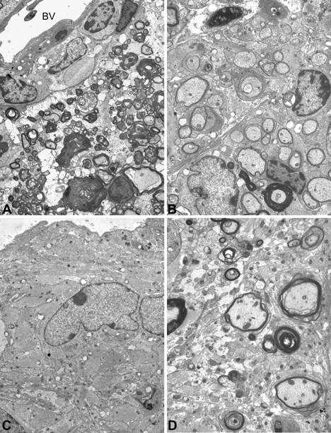
Activation of the classical complement system is known to play a central role in autoimmune demyelination. We have analyzed the role of complement component C5 in experimental autoimmune encephalomyelitis (EAE) using C5-deficient (C5-d) and C5-sufficient (C5-s) mice. Both groups of mice displayed early onset EAE, a short recovery phase, and similar stable chronic courses. However, in contrast to the clinical similarities, marked differences were apparent by histopathology. During acute EAE in C5-d, a delay in inflammatory cell infiltration and tissue damage was observed along with restricted lesion areas, which in C5-s mice were more extensive and diffuse. More striking were the differences in chronic lesions. In C5-d mice, inflammatory demyelination and Wallerian degeneration were followed by axonal depletion and severe gliosis, while in C5-s, the same initial signs were followed by axonal sparing and extensive remyelination. In C5-d, immunohistochemistry and Western blotting showed an increase in glial fibrillary acidic protein and a decrease in neurofilament protein, proteolipid protein, and several pro-inflammatory markers. These results in the EAE model indicate that absence of C5 resulted in fiber loss and extensive scarring, whereas presence of C5-favored axonal survival and more efficient remyelination.


Figures

Figure 1.
Figure 1.
Serum hemolytic activity and C5 mRNA expression in C5-d mice. A: Mouse serum was tested for the ability of serum complement to lyse rabbit erythrocytes by the alternative pathway. Results are expressed by determining the released hemoglobulin at OD412 nm. Control C5-s serum had a hemolytic activity of 2.5 (result not shown). The mean hemolytic activity of five C5-d mice with acute EAE was very low, compared to the mean activity of four C5-s mice. These findings confirmed the lack of activation and consumption of complement during acute EAE in C5-d mice. B: RNA obtained from C5-d and C5-s spinal cords and brain from one C5-s mouse was analyzed for C5 mRNA expression by RT-PCR. Analysis of the RT-PCR products on a 1% agarose gel demonstrated only in C5-s mice a band of 400 bp using C5-specific primer.
Figure 2.
Figure 2.
Summary of clinical scores of C5-d and C5-s mice with EAE. The disease pattern of 80 C5-d (B10D2/oSnJ) and 80 C5-s (B10.D2/nSnJ) outbred mice is shown as the mean ± SD. After an initial peak and a recovery period, the animals developed a stable chronic form of EAE, which in C5-s mice was slowly progressive. 75% of C5-d mice and 96% of C5-s mice developed a disease score of 2.5 to 3.5 between days 10 and 11 p.i. Animals were sampled at the onset of disease phase (9 to 14 days p.i.; 23 C5-d, 24 C5-s mice), acute phase (17 to 19 days p.i.; 3 mice each), recovery phase (21 to 25 days p.i.; 16 C5-d, 23 C5-s) or chronic phase [50 (2 C5-d, 3 C5-s), 90 (2 C5-d, 4 C5-s), 117 and 120 days p.i. (20 C5-d, 18 C5-s)].
Figure 3.
Figure 3.
Histopathology of acute, recovery, and chronic phases of EAE in C5-d and C5-s mice. Toluidine-blue-stained epoxy sections (1 μm) from the upper lumbar of C5-d mice with acute EAE (A and B), revealed well-demarcated, confined lesions with inflammation and a narrow rim of demyelinated axons. Lesions of C5-s mice with acute EAE (C and D) are more diffuse and display inflammation and demyelination in addition to Wallerian degeneration (D, lower left), dystrophic axons (D, center right), and myelin ovoids (D, left). In the recovery phase of EAE, C5-d mice (E and F, cervical spinal cord) show extensive Wallerian degeneration (dense droplets, lower right) and demyelination. In contrast, extensive remyelination was present in C5-s mice (G and H, S1 spinal cord), shown by numerous thinly myelinated fibers along the meningeal surface (H). Numerous fibrous astrocytes with pale nuclei occur within the remyelinated zone. During chronic EAE, C5-d mice (I and J) developed a prominent zone of intense gliosis at the margin of the spinal cord, from which nerve fibers and oligodendrocytes were depleted. Lesions in C5-s mice (K and L) displayed numerous remyelinated axons at the edge of the spinal cord, where some gliosis is also present.
Figure 4.
Figure 4.
Immunohistochemistry (IH) of C5-d and C5-s mice with EAE. Neurofilament (NF-200): Sections from C5-d with chronic EAE revealed weaker and scattered staining of NF-200 (A) in areas corresponding to a chronic lesion, compared to the robust, densely-packed staining in sections of C5-s mice (B), where most axons survived. P-Selectin: In chronic EAE, staining for the endothelial adhesion molecule, P-Selectin, was lower in C5-d (C) than C5-s (D), perhaps indicating reduced infiltration of inflammatory cells into the tissue. MAC-1: Increased staining of macrophages in C5-s mice (E), during acute disease suggested enhanced phagocytosis of myelin debris in C5-s, in comparison to C5-d mice (F). IL-6: In acute EAE, IL-6 was enhanced in reactive astrocytes in C5-d (G), in comparison to C5-s mice (H). Magnifications: A and B, ×63; E, F, G, H, ×625.
Figure 5.
Figure 5.
Protein expression by Western blot analysis. 10 μg (GFAP, PLP, and C1q) or 100 μg (VCAM-1 and TNF-α) of spinal cord homogenate separated on 10% to 12% SDS-PAGE and transferred by Western blotting were probed with specific antibodies for various cytokines, adhesion molecules and myelin proteins. Results performed in triplicate (except for PLP, VCAM-1) are expressed as relative values of densitometric numbers with the value of the band obtained for untreated C5-s spinal cord taken as 1.0. GFAP: GFAP (A) up-regulated by IH during the chronic phase in C5-d in comparison to C5-s mice was confirmed, indicating enhanced astrogliosis in C5-d. PLP: The myelin protein, PLP (B), was shown to be down-regulated during the acute phase in both groups, indicating myelin breakdown. In chronic C5-s mice, almost baseline levels of PLP were detected, suggesting remyelination. C1q: In C5-d mice, the complement protein, C1q (C), was decreased in comparison to C5-s during acute and even more so during chronic EAE, indicating an increased activation of complement in C5-s mice. VCAM-1: A robust inflammatory response, indicated by up-regulation of the adhesion molecule, VCAM-1 (D), during the acute phase, was sustained further in C5-d mice during chronic disease, reaching levels similar to levels in chronic C5-s. TNF-α: Up-regulation of TNF-α was more prominent in C5-d mice than C5-s during acute EAE, whereas levels dropped in C5-d mice but not C5-s mice during chronic EAE (P < 0·026) (E).
Figure 6.
Figure 6.
Differences between chronic lesions in C5-d and C5-s mice by EM. An area of a spinal cord from a C5-d mouse (A, 24 days p.i.) shows destructive lesions with ongoing Wallerian degeneration and a few inflammatory cells, whereas in C5-s (B, 24 days p.i.) widespread early remyelination is clearly visible. During chronic EAE (C, 120 days p.i.), differences were equally obvious, with fibrous astrogliosis predominating in C5-d mice, and advanced remyelination and a less gliotic parenchyma in matching sections of C5-s mice (D). Magnifications: A, ×3400; B, ×4600; C, ×5400; D, ×6000.

Similar articles

Cited by

References

    1. Botto M: C1q knockout mice for the study of complement deficiency in autoimmune disease. Exp Clin Immunogenet 1998, 15:231-234 - PubMed
    1. Wessels MR, Butko P, Ma M, Warren HB, Lage AL, Carroll MC: Studies of group B streptococcal infection in mice deficient in complement component C3 or C4 demonstrate an essential role for complement in both innate and acquired immunity. Proc Natl Acad Sci USA 1995, 92:11490-11494 - PMC - PubMed
    1. Morgan BP, Walport MJ: Complement deficiency and disease. Immunol Today 1991, 12:301-306 - PubMed
    1. Gasque P, Dean YD, McGreal EP, VanBeek J, Morgan BP: Complement components of the innate immune system in health and disease in the CNS. Immunopharmacology 2000, 49:171-186 - PubMed
    1. Shin M, Rus H, Niculescu F: Complement system in central nervous system disorders. Volankis JE Frank MM eds. The Human Complement in Health and Disease 1998:pp 499-525 Marcel Dekker, New York

Publication types

MeSH terms