. 2003 Nov;14(11):4569-80.
doi: 10.1091/mbc.e03-01-0009. Epub 2003 Aug 22.

Matrix metalloproteinase 19 regulates insulin-like growth factor-mediated proliferation, migration, and adhesion in human keratinocytes through proteolysis of insulin-like growth factor binding protein-3

Affiliations

Matrix metalloproteinase 19 regulates insulin-like growth factor-mediated proliferation, migration, and adhesion in human keratinocytes through proteolysis of insulin-like growth factor binding protein-3

Thorsten Sadowski et al. Mol Biol Cell. 2003 Nov.

Abstract

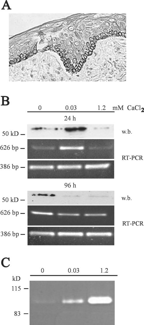
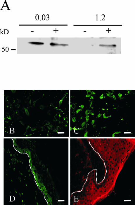
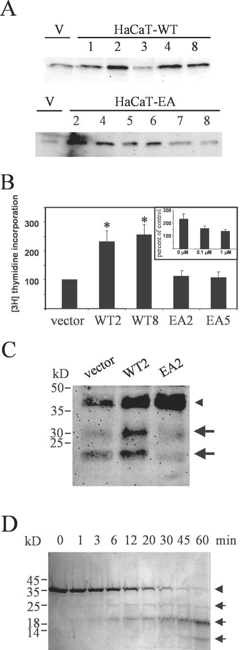
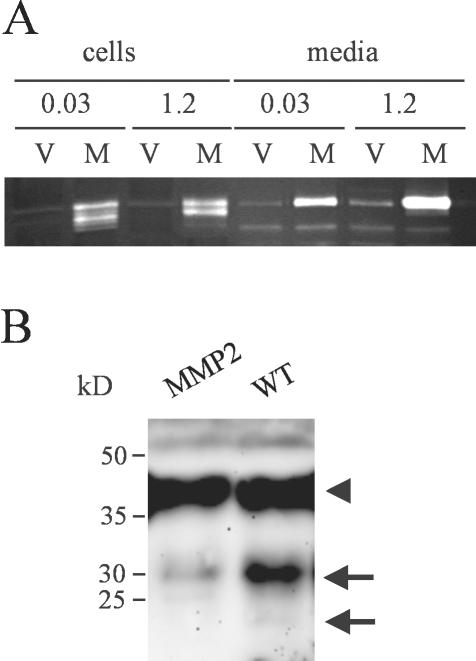
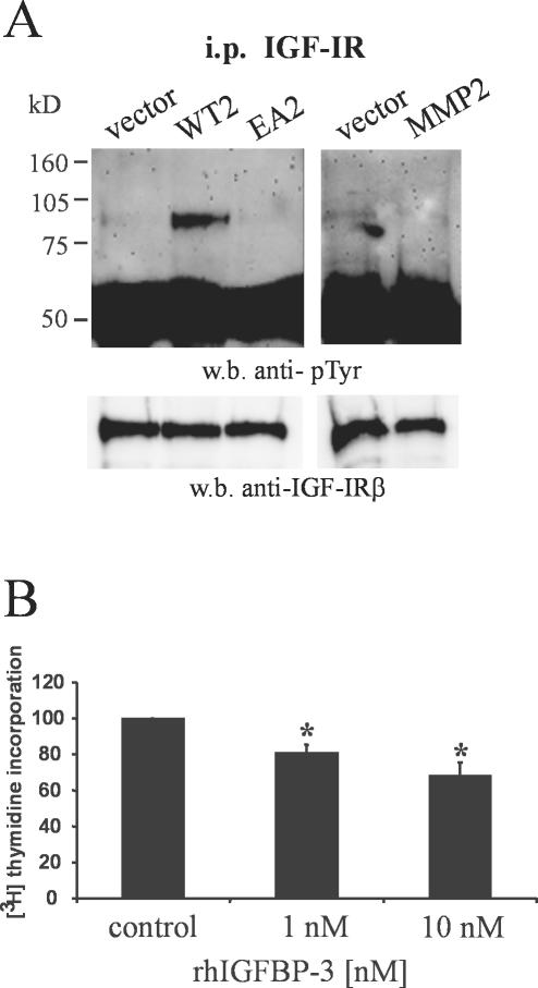
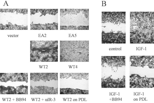
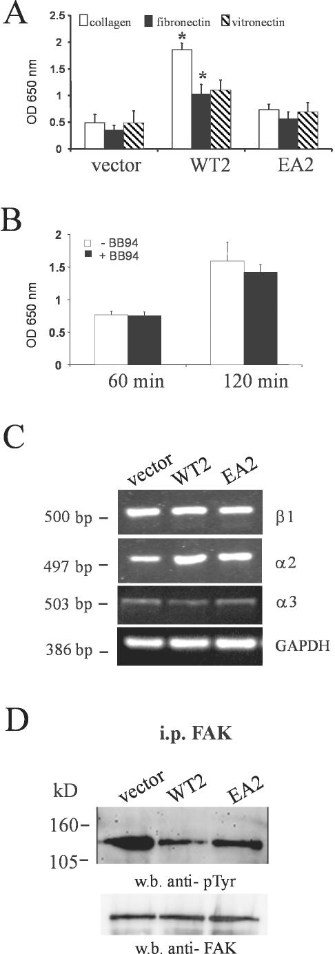
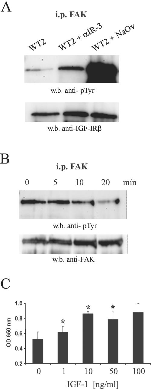
Unlike most other matrix metalloproteinases (MMPs) MMP-19 is expressed in undifferentiated basal keratinocytes of healthy human skin. The human keratinocyte cell line HaCaT, which like basal keratinocytes constitutively expresses MMP-19, down-regulated the expression of MMP-19 at high calcium concentrations. Calcium-regulation occurred through E-cadherin mediated cell-cell contacts because neutralizing anti-E-cadherin antibodies restored MMP-19 expression in high calcium. Overexpression of MMP-19 in HaCaT cells (HaCaT-WT) increased cellular proliferation, as well as migration and adhesion on type I collagen. This was due to proteolysis of the insulin-like growth factor (IGF) binding protein-3 by MMP-19, which augmented signaling through the IGF-I receptor, as evidenced by its increased autophosphorylation. Conversely, these effects were not observed in cells transfected with MMP-2 or a catalytically inactive MMP-19 mutant. As further proof that increased IGF-signaling promoted adhesion and migration in HaCaT-WT cells, we reproduced these effects by treating parental HaCaT with IGF-I. We observed dephosphorylation of the focal adhesion kinase in HaCaT-WT as well as IGF-I-treated HaCaT cells, suggesting that inactivating focal adhesion kinase is a mechanism by which IGF-I enhances adhesion. Furthermore, IGF-I-triggered motility on type I collagen was mediated by MMP activity, which, however, was distinct from MMP-19. Considering the coexpression of IGFBP-3 and MMP-19 in the skin, we conclude that MMP-19 is a likely candidate to be the major IGFBP-3 degrading MMP in the quiescent epidermis. This activity might have widespread consequences for the behavior of epidermal keratinocytes.


Figures

Figure 1.
Figure 1.
MMP-19 expression in keratinocytes is dependent on cellular differentiation. (A) Sections of paraffin-embedded samples of human skin with normal morphology were analyzed with antibodies against MMP-19. The mAb CK8/4 detects MMP-19 specifically in basal keratinocytes. Bars, 50 μm. (B) MMP-19 protein and mRNA expression of HaCaT after 24 h and 96 h in keratinocyte-SFM with 0, 0.03, and 1.2 mM calcium. Cell lysates were subjected to Western blotting (w.b.) and probed with polyclonal antibodies against human MMP-19. A specific signal was detected at the expected size of ∼57 kDa (top). For evaluating the mRNA expression of MMP-19, total RNA was isolated and analyzed by RT-PCR with MMP-19-specific primers (middle). GAPDH was used as an internal control (bottom). Results are representative of three experiments. (C) The proform of MMP-9 is detected in conditioned media of HaCaT grown for 96 h in keratinocyte-SFM with 0, 0.03, and 1.2 mM calcium. Shown is a gelatinolytic zymogram. Results are representative of three experiments.
Figure 2.
Figure 2.
Suppression of MMP-19 expression by high calcium is mediated by cell-cell contacts. (A) Cytochalasin D (5 μg/ml) was used to disrupt adherens junctions. HaCaT keratinocytes were grown for 24 h in keratinocyte-SFM containing 0.03 and 1.2 mM calcium. Lysates of cytochalasin-treated (+) and untreated (-) cells were subjected to Western blotting for MMP-19. (B and C) The E-cadherin–specific antibody DECMA-1 down-regulated the expression of MMP-19. The DECMA-1 antibody (C) and an isotype control (B) were added at a final concentration of 1:15. HaCaT keratinocytes were grown in keratinocyte-SFM containing 1.2 mM calcium for 24 h. Afterwards, cells were fixed in 4% paraformaldehyde and processed for immunofluorescent detection of MMP-19 as described in Materials and Methods. Bars, 50 μm. The image shown is representative of three independent experiments. (C and D) Frozen sections of normal human epidermis were analyzed with antibodies against MMP-19 (C) and E-cadherin (D). Immunofluorescent detection was by Alexa 488 (MMP-19) and Cy 3 (E-cadherin)-conjugated secondary antibodies. Dotted lines indicate the basement membrane. Bars, 50 μm. Note that the expression of these molecules localizes to distinct compartments of human epidermis.
Figure 3.
Figure 3.
Increased proliferation and proteolysis of IGFBP-3 in keratinocytes overexpressing wild-type MMP19. (A) Several clones of HaCaT transfected with plasmids coding for the wild-type (HaCaT-WT) and for a functional knockout mutant of MMP-19 (HaCaT-EA) were assayed for overexpression of MMP-19 by comparison with HaCaT carrying the empty vector (V) by using Western blotting. (B) Independent clones of HaCaT-WT (WT), HaCaT-EA (EA), and HaCaT-pIRES (vector) were seeded at 15,000 cells/well in microtiter plates and grown for 24 h in standard culture medium. At the end of the incubation period, cells were pulsed with 0.25 μCi/well [3H]thymidine for an additional 4 h. Values obtained for cells carrying the empty vector were considered 100%. Given are the means ± SEM (n = 3). *, significantly different from HaCaT-pIRES with p ≤ 0.05. Inset, effect of the hydroxamate MMP-inhibitor BB94 on the proliferation of HaCaT-WT2 cells. (C) HaCaT-WT2 (WT2), HaCaT-EA2 (EA2), and HaCaT-pIRES (vector) were grown for 72 h in keratinocyte-SFM. Conditioned media were concentrated 10-fold and analyzed for IGFBP-3 proteolysis by Western blotting. The arrowhead indicates the position of intact IGFBP-3, whereas arrows point to 30- and 19-kDa IGFBP-3 proteolytic fragments. This experiment is representative of three independent experiments. (D) rhIGFBP-3 (240 μg/ml) was incubated with GST-MMP-19 (300 ng/ml) in TNC buffer (50 mM Tris, 150 mM NaCl, 10 mM CaCl2, 0.05% Brij 35, 20 μM ZnCl2, pH 7.5) at 37°C. Before starting the reaction, a 10-μl sample was taken, whereas the reaction-tube was still on ice, representing the intact IGFBP-3. At the indicated time points, samples were taken and immediately mixed with Laemmli buffer. The samples were run on a 12% SDS-polyacrylamide gel under reducing conditions. The arrowhead indicates the position of intact IGFBP-3, whereas arrows point to IGFBP-3 proteolytic fragments.
Figure 4.
Figure 4.
MMP-2 does not contribute to the degradation of IGFBP-3 in HaCaT keratinocytes. (A) Conditioned media and lysates of HaCaT-MMP2 (M) grown for 3 d in keratinocyte-SFM with 0.03 mM and 1.2 mM calcium were analyzed for overexpression of MMP-2 by comparison with HaCaT carrying the empty vector (V) by using gelatin zymography. Although media contained only the MMP-2 proform, processed MMP-2 was found to be associated with the cells. (B) HaCaT-WT (WT) and HaCaT-MMP2 (MMP2) were grown for 72 h in keratinocyte-SFM. Conditioned media were concentrated 10-fold and analyzed for IGFBP-3 proteolysis by Western blotting. The arrowhead indicates the position of intact IGFBP-3, whereas arrows point to 30- and 19-kDa IGFBP-3 proteolytic fragments. This experiment is representative of three independent experiments.
Figure 5.
Figure 5.
Wild-type MMP-19 increases IGF-triggered cell signaling in keratinocytes. (A) IGF-IR was immunoprecipitated from HaCaT-WT2 (WT2), HaCaT-EA2 (EA2), HaCaT-MMP2 (MMP2), and HaCaT-pIRES (vector). Precipitates were immunoblotted and membranes were probed sequentially with anti-phosphotyrosine and polyclonal anti-IGF-IR antibodies. (B) HaCaT-WT2 cells were seeded at 15,000 cells/well in microtiter plates and grown for 48 h in standard culture medium in the absence (control) or presence of 1 and 10 nM rhIGFBP-3. Values obtained without rhIGFBP-3 were considered 100%. Given are the means + SEM (n = 3). *, significantly different from control with p < 0.05.
Figure 6.
Figure 6.
MMP-19 regulates IGF-stimulated migration on type I collagen. (A) Independent clones of HaCaT-WT (WT), HaCaT-EA (EA), and HaCaT-pIRES (vector) were seeded on type I collagen. At confluence, a cell-free area was introduced and migration was evaluated after 48 h. Recovery of the denuded area by HaCaT-WT2 was inhibited in the presence of 1 μM BB94 and 1 μg/ml anti-IGF-IR antibody (αIR3), and did not occur when cells were seeded on the nonintegrin substrate poly-d-lysine (PDL). (B) HaCaT cells were seeded on type I collagen. At confluence, a cell-free area was introduced and cells were left untreated (control) or treated with 100 ng/ml IGF-I in the presence or absence of 1 μM BB94. In addition, migration of HaCaT was evaluated on PDL in the presence of 100 ng/ml IGF-I.
Figure 7.
Figure 7.
Increased adhesion of HaCaT-WT cells is not dependent on altered integrin expression but correlates with dephosphorylation of FAK. (A) HaCaT-WT2 (WT2), HaCaT-EA2 (EA2), and HaCaT-pIRES (vector) were seeded on type I collagen, vitronectin, and fibronectin matrices and were allowed to adhere for 2 h. Nonadherent cells were then washed away and attached cells were quantitated as described under MATERIALS AND METHODS. Given are the means + SEM (n = 3). *, significantly different from HaCaT-pIRES with p <0.05. (B) Time course of HaCaT-WT2 adhesion to type I collagen after pretreatment of the cells with BB94 (1 μM). (C) For evaluating the mRNA expression of integrins in HaCaT-WT2 (WT2), HaCaT-EA2 (EA2), and HaCaT-pIRES (vector), total RNA was isolated and analyzed by RT-PCR with primers specific for β1, α2, and α3 integrin. GAPDH was used as an internal control. Results are representative of three experiments. (D) FAK was immunoprecipitated from HaCaT-WT2 (WT2), HaCaT-EA2 (EA2), and HaCaT-pIRES (vector). Precipitates were immunoblotted and membranes were probed sequentially with anti-phosphotyrosine and polyclonal anti-FAK antibodies.
Figure 8.
Figure 8.
MMP-19–induced activation of the IGF-IR–mediated dephosphorylation of FAK and increased adhesion to type I collagen. (A) HaCaT-WT2 cells (WT2) were incubated for 24 h with 1 μg/ml anti-IGF-IR antibody (αIR-3) or 100 nM sodium orthovanadate (NaOv). FAK was immunoprecipitated from the lysates and precipitates were immunoblotted. Membranes were probed sequentially with anti-phosphotyrosine and polyclonal anti-FAK antibodies. (B) HaCaT cells were serum starved for 24 h. IGF-I (100 ng/ml) was added and incubated for the indicated time points. FAK was immunoprecipitated from the lysates and analyzed for phosphorylation as described above. (C) HaCaT was incubated with the indicated concentrations of IGF-I for 24 h under serum-free conditions. Cells were then trypsinized and seeded on type I collagen and were allowed to adhere for 2 h. Nonadherent cells were then washed away and attached cells were quantified as described under MATERIALS AND METHODS. Given are the means + SEM (n = 3). *, significantly different from control with p < 0.05.

Similar articles

Cited by

References

    1. Ando, Y., and Jensen, P.J. (1993). Epidermal growth factor and insulin-like growth factor I enhance keratinocyte migration. J. Investig. Dermatol. 100, 633-639. - PubMed
    1. Baron, V., Calleja, V., Ferrari, P., Alengrin, F., and van Obberghen, E. (1998). p125FAK focal adhesion kinase is a substrate for the insulin and insulin-like growth factor-I tryrosine kinase receptors. J. Biol. Chem. 273, 7162-7168. - PubMed
    1. Bikle, D.D., and Pillai, S. (1993). Vitamin D, calcium, and epidermal differentiation. Endocr. Rev. 14, 3-19. - PubMed
    1. Birkedahl-Hansen, H., Moore, W.G., Bodden, M.-K., Windsor, L.J., Birkedahl-Hansen, B., DeCarlo, A., and Engler, J.-A. (1993). Matrix metalloproteinases: a review. Crit. Rev. Oral Biol. Med. 4, 197. - PubMed
    1. Brabletz, T., Jung, A., Dag, S., Hlubek, F., and Kirchner, T. (1999). beta-Catenin regulates the expression of the matrix metalloproteinase-7 in human colorectal cancer. Am. J. Pathol. 155, 1033-1038. - PMC - PubMed

Publication types

MeSH terms