. 2003 Nov;14(11):4541-56.
doi: 10.1091/mbc.e03-02-0078. Epub 2003 Aug 22.

Inhibition of proteasomal activity causes inclusion formation in neuronal and non-neuronal cells overexpressing Parkin

Affiliations

Inhibition of proteasomal activity causes inclusion formation in neuronal and non-neuronal cells overexpressing Parkin

Helen C Ardley et al. Mol Biol Cell. 2003 Nov.

Abstract

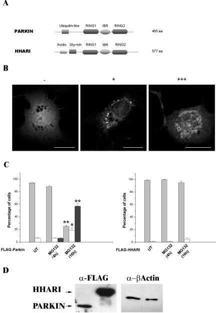
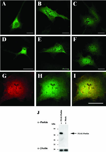
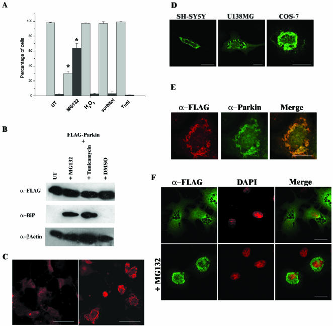
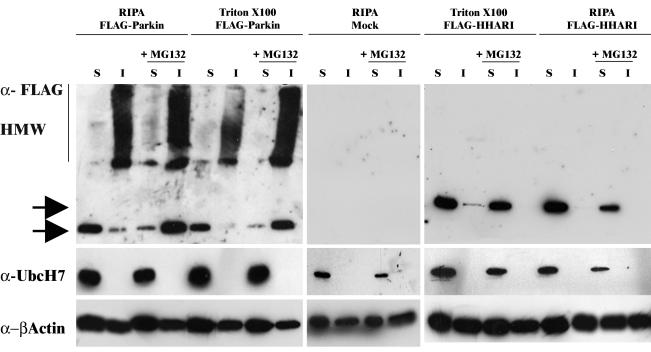
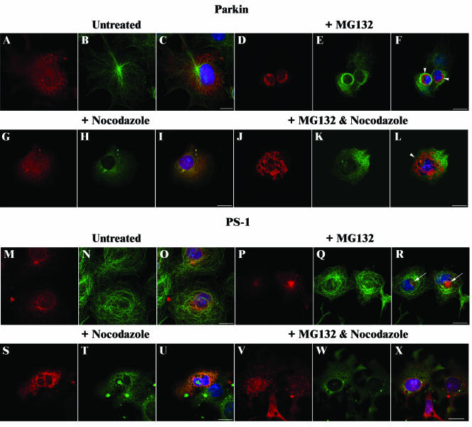
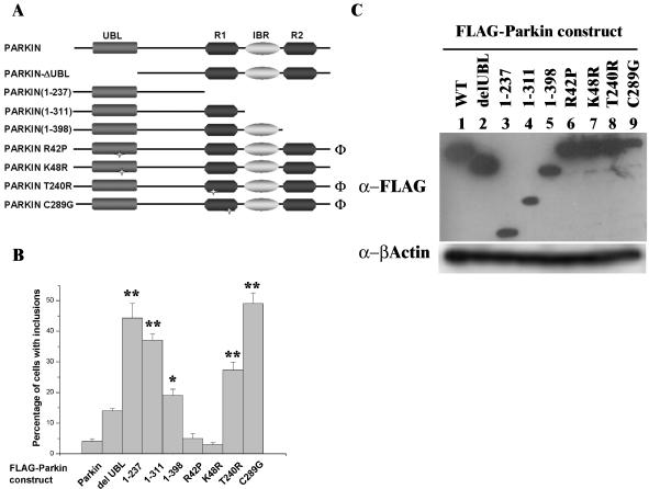
Association between protein inclusions and neurodegenerative diseases, including Parkinson's and Alzheimer's diseases, and polyglutamine disorders, has been widely documented. Although ubiquitin is conjugated to many of these aggregated proteins, the 26S proteasome does not efficiently degrade them. Mutations in the ubiquitin-protein ligase Parkin are associated with autosomal recessive juvenile Parkinsonism. Although Parkin-positive inclusions are not detected in brains of autosomal recessive juvenile Parkinsonism patients, Parkin is found in Lewy bodies in sporadic disease. This suggests that loss of Parkin ligase activity via mutation, or sequestration to Lewy bodies, is a contributory factor to sporadic disease onset. We now demonstrate that decreased proteasomal activity causes formation of large, noncytotoxic inclusions within the cytoplasm of both neuronal and nonneuronal cells overexpressing Parkin. This is not a general phenomenon as there is an absence of similar inclusions when HHARI, a structural homolog of Parkin, is overexpressed. The inclusions colocalize with ubiquitin and with proteasomes. Furthermore, Parkin inclusions colocalize with gamma-tubulin, acetylated alpha-tubulin, and cause redistribution of vimentin, suggesting aggresome-like properties. Our data imply that lower proteasomal activity, previously observed in brain tissue of Parkinson's disease patients, leads to Parkin accumulation and a concomitant reduction in ligase activity, thereby promoting Lewy body formation.


Figures

Figure 3.
Figure 3.
Inclusion body formation by Parkin is not a general response of RING-IBR-RING proteins to proteasome inhibition. (A) Schematic illustrating the domain structures of Parkin and HHARI. Parkin and HHARI demonstrate structural homology at the 3′ C-terminus. (B) COS-7 cells transfected with the FLAG-Parkin construct, stained with anti-FLAG antibodies as described for Figure 1. Cells were scored as either containing no inclusions (-), small individual inclusions (+), or large inclusions (+++). Bars, 20 μm. (C) COS-7 cells were transfected with FLAG-Parkin (left) or FLAG-HHARI (right). MG132 (20 μM) was added to cells for 4 or 16 h before fixation. The presence of Parkin or HHARI was assessed by immunofluorescence using the anti-FLAG antibody as described for Figure 1. Light gray bars, -; white bars, +; and dark gray bars, +++. Error bars indicate the SE from the mean. The asterisk (*) indicates a significant difference between the percentage of untreated cells with or without inclusions vs. the numbers of MG132-treated cells at each time point. *p < 0.005; ** p < 0.001. N-Terminal Myc-tagged and pDsRed1-C1 Parkin or HHARI constructs produced similar results. (D) Lack of HHARI inclusions is not due to poor expression of the construct. COS-7 cells were transfected with FLAG-Parkin or FLAG-HHARI. Forty-four hours post-transfection, cell lysates were prepared in Triton X-100 buffer. Ten micrograms of each soluble fraction was separated by SDS-PAGE and analyzed by Western blotting with anti-FLAG and anti-β-actin antibodies. Arrows indicate the presence of the respective FLAG construct.
Figure 1.
Figure 1.
Localization of Parkin in neuronal and nonneuronal cell lines. FLAG-Parkin was transfected into SH-SY5Y (A and D), U138MG (B and E), or COS-7 (C and F–I). Forty-four hours post-transfection, the cells were fixed with 4% (wt/vol) paraformaldehyde, permeabilized, and immunostained with mouse monoclonal anti-FLAG (1:1000) (A–C and G), or affinity-purified rabbit anti-Parkin peptide (1:50) (D–F and H) antibodies. Anti-FLAG and anti-Parkin demonstrate overlapping staining patterns in COS-7 cells transfected with FLAG-Parkin (I, yellow staining indicates regions of colocalization within cells). Bars, 20 μm. (J) COS-7 cells lysates from FLAG-Parkin (lane 1) or mock (lane 2)-transfected cells were prepared in RIPA buffer 48 h post-transfection. Ten micrograms of each total cell lysate was separated by SDS-PAGE and analyzed by Western blotting with affinity-purified anti-Parkn C-terminal peptide (1: 500) and anti-β-actin (1:5000) antibodies. Arrow indicates the presence of FLAG-Parkin. A band corresponding to the expected position of endogenous Parkin was not observed in either lane.
Figure 2.
Figure 2.
Addition of the proteasomal inhibitor MG132 causes inclusion body formation in cells overexpressing Parkin. (A) COS-7 cells were transfected with FLAG-Parkin. Twenty-eight hours post-transfection, cells were incubated for 16 h in the presence of either 20 μM MG132, 400 μM hydrogen peroxide (H2O2), 300 mM sorbitol, or 10 μg/ml tunicamycin. The presence of Parkin was then assessed by immunofluorescence with mouse monoclonal anti-FLAG antibody. Light gray bars represent the percentage of cells demonstrating a “wild-type” staining pattern, and dark gray bars represent the percentage of cells containing inclusion bodies. A minimum of 300 transfected cells was analyzed for each sample. Data shown represent the results of two independent sets of experiments. Error bars indicate the SE from the mean of these experiments. The asterisk (*) indicates a significant difference between the percentage of untreated cells versus the percentage of treated cells. *p < 0.01. (B) Tunicamycin and the proteasome inhibitor MG132 induce the UPR. COS-7 cell lysates from FLAG-Parkin–transfected cells left untreated (UT) or treated with 20 μM MG132, 10 μg/ml tunicamycin, or the carrier DMSO for 16 h were prepared in RIPA buffer 48 h post-transfection. Ten micrograms of each total cell lysate was separated by SDS-PAGE and analyzed by Western blotting with anti-FLAG, anti-BiP (1:250), and anti-β-actin antibodies. (C) COS-7 cells were transfected with FLAG-Parkin. Twenty-eight hours post-transfection, cells were grown in the presence or absence of 20 μM MG132 for 16 h before fixation. The presence of Parkin was assessed as described above. COS-7 cells containing MG132 induced inclusions (right) display a more intense staining pattern than untreated cells overexpressing Parkin (left). Confocal images of cells were scanned at the same fluorescent intensity. Bars, 50 μm. (D) Proteasome inhibition induces inclusion formation in SH-SY5Y, U138MG and COS-7 cells overexpressing FLAG-Parkin. (E) Immunostaining of inclusions with anti-FLAG (red) or anti-Parkin peptide (green) antibodies demonstrated similar staining patterns as indicated by yellow staining in merged panel. (F) Inclusions formed are within the cytosol. Transfected COS-7 cells were assessed by immunofluorescence by using the monoclonal anti-FLAG antibodies (green) and counterstained with 4,6-diamidino-2-phenylindole to visualize nuclei (red). Bars (D–F), 20 μm.
Figure 4.
Figure 4.
MG132 induces the formation of insoluble, high-molecular-weight Parkin species. COS-7 cells were transfected with FLAG-Parkin (left), FLAG-HHARI (right), or mock-transfected (middle) and grown in the presence or absence of MG132 for 16 or 28 h post-transfection. Cell lysates were prepared in buffer as indicated, and 10 μg of each soluble (S) and insoluble (I) fractions was separated by SDS-PAGE and analyzed by Western blotting. Blots were probed with mouse anti-FLAG (top), affinity-purified rabbit anti-UbcH7 peptide (1:5000) (middle), and anti-β-actin (bottom) antibodies. Arrows indicate the presence of each respective FLAG construct.
Figure 5.
Figure 5.
Effect of nocodazole on Parkin and PS-1 inclusions. COS-7 cells were transfected with FLAG-Parkin (A–L) or PS1 (M–X) constructs and grown in the absence (A–C and M–O) or presence of 20 μM MG132 (D–F and P–R) or 10 μg/ml nocodazole (G–I and S–U) or both (J–L and V–X) for 16 h. After methanol fixation, cells were probed with affinity-purified rabbit anti-Parkin peptide (1:50) (A–L; red), or affinity-purified rabbit anti-PS1 peptide (1:50) (M–X; red) antibodies, and rat anti-α-tubulin (1:500) (A–X; green) antibodies. Overlays of each set of three include 4,6-diamidino-2-phenylindole staining (blue) to identify nuclei (C, F, I, L, O, R, U, and X). Regions of colocalization within cells stain yellow. Arrowheads indicate Parkin inclusions; arrows indicate PS1 aggresomes. Bars, 20 μm.
Figure 6.
Figure 6.
Parkin inclusion bodies display aggresome-like characteristics. (A–H) Parkin inclusions cause disruption of γ-tubulin localization. COS-7 cells were transfected with FLAG-Parkin (A–C and E–G) in the presence (E–H) or absence (A–D) of MG132. Cells were stained with affinity-purified rabbit anti-Parkin (red) and mouse γ-tubulin (1:1000) (green) antibodies. Staining of untransfected cells confirmed aggregation of γ-tubulin only occurs in cells overexpressing Parkin (D and H). Red staining in D and H represents 4,6-diamidino-2-phenylindole staining of the nucleus. Arrowheads indicate γ-tubulin localization. (I–N) Partial colocalization of Parkin and vimentin near the centrosome. Cells overexpressing Parkin were stained with affinity-purified rabbit anti-Parkin (red) and anti-vimentin (1:40) antibodies (green) in the presence (L–N) or absence (I–K) of MG132. Arrow indicates region of colocalization in N. (O–Q) Acetylated α-tubulin localizes to Parkin inclusions. Cells overexpressing Parkin were stained with affinity-purified rabbit anti-Parkin (red) and mouse anti-acetylated α-tubulin (1:2000) antibodies (green). Panels show untreated (O), MG132-treated (P), or combined MG132 and nocodazole-treated (Q) cells. Arrows indicate colocalization within the inclusion in P and Q. The asterisk (*) indicates an untransfected cell with intact acetylated α-tubulin staining in Q. Overlays of each set include 4,6-diamidino-2-phenylindole staining (blue) (C, G, K, and N) to identify the nucleus. Mitochondria localize with Parkin inclusions. In R and S, Parkin expression is indicated by the green fluorescence and the mitochondria were labeled using MitoTracker (used at 300 nM; red fluorescence). With the exception of (R and S), which were fixed with 4% (wt/vol) paraformaldehyde, all cells were fixed with methanol. Bars, 20 μm.
Figure 7.
Figure 7.
Colocalization of Parkin inclusions with other components of the ubiquitin-proteasome pathway. (A–R) COS-7 cells were transfected with the FLAG-Parkin expression construct and cultured in the presence or absence of MG132 for 16 h. Cells were probed with the following: A–F, anti-FLAG and affinity-purified rabbit anti-UbcH7 peptide (1:100) antibodies; ubiquitin (1:50) antibodies; G–L, anti-FLAG and anti-20S (1:100) antibodies; or M–R, anti-FLAG and anti-ubiquitin (1:50) antibodies. Staining was as follows: left, FLAG, to identify Parkin (red); middle, UbcH7 (B and E), 20S (H and K), or ubiquitin (N and Q) (green); and right, overlay of each set with the addition of 4,6-diamidino-2-phenylindole staining (blue) to identify the nucleus. Regions of colocalization within cells stain yellow. Arrows indicate colocalization within an inclusion. Bars, 20 μm. (S) COS-7 cells were transfected with the Myc-Parkin expression construct alone or cotransfected with the FLAG-tagged ubiquitin construct as indicated and cultured in the absence of MG132. Cells were analyzed by immunofluorescence by using anti-Parkin antibodies as described for Figure 1 and assessed as described in Figure 3. Error bars indicate the SE from the mean. The asterisk (*) indicates a significant difference between the number of Myc-Parkin/FLAG-ubiquitin construct-transfected cells containing inclusions versus the number found in Myc-Parkin alone transfected cells; p < 0.01. Costaining with anti-FLAG antibody revealed that exogenous ubiquitin had an identical staining pattern to the endogenous protein (our unpublished data).
Figure 8.
Figure 8.
Effects of truncation and point mutations of Parkin on inclusion body formation. (A) Schematic illustrating the positions of the point mutations and truncations of Parkin constructs used in B. Φ, AR-JP–associated mutation. (B) COS-7 cells were transfected with FLAG-Parkin or deletion/mutation constructs and cultured in the absence of MG132. The expression of Parkin was analyzed by immunofluorescence with anti-FLAG antibodies. Cells were scored for the presence of inclusion bodies. Error bars indicate the SE from the mean. Asterisk(s) indicate a significant difference between the number of inclusions observed with each construct compared with wild type, FLAG-Parkin, without addition of MG132. *p < 0.05; **p < 0.001. (C) Inclusion formation is not related to expression levels of the Parkin constructs. COS-7 cells were transfected with each FLAG-Parkin construct as indicated. Forty-four hours post-transfection, cell lysates were prepared in RIPA buffer. Ten micrograms of total cell lysate was separated by SDS-PAGE and analyzed by Western blotting with anti-FLAG (top) and anti-β-actin (bottom) antibodies.

Similar articles

Cited by

References

    1. Apostol, R.L., et al. (2003). A cell-based assay for aggregation inhibitors as therapeutics of polyglutamine-repeat disease and validation in Drosophila. Proc. Natl. Acad. Sci. USA 100, 5950-5955. - PMC - PubMed
    1. Ardley, H.C., Tan, N.G.S., Rose, S.A., Markham, A.F., and Robinson, P.A. (2001). Features of the Parkin/Ariadne-like ubiquitin ligase, HHARI, that regulate its interaction with the ubiquitin-conjugating enzyme, UbcH7. J. Biol. Chem. 276, 19640-19647. - PubMed
    1. Arima, K., Mizutani, T., Alim, M.A., Tonozuka-Uehara, H., Izumiyama, Y., Hirai, S., and Ueda, K. (2000). NACP/alpha-synuclein and tau constitute two distinctive subsets of filaments in the same neuronal inclusions in brains from a family of parkinsonism and dementia with Lewy bodies: double-immunolabeling fluorescence and electron microscopic studies. Acta Neuropathol. 100, 115-121. - PubMed
    1. Baas, P.W., and Black, M.M. (1990). Individual microtubules in the axon consist of domains that differ in both composition and stability. J. Cell Biol. 111, 495-509. - PMC - PubMed
    1. Bence, N.F., Sampat, R.M., and Kopito, R.R. (2001). Impairment of the ubiquitin-proteasome system by protein aggregation. Science 292, 1552-1555. - PubMed

Publication types

MeSH terms