. 2003 Nov;14(11):4641-53.
doi: 10.1091/mbc.e03-02-0091. Epub 2003 Aug 22.

Villin enhances hepatocyte growth factor-induced actin cytoskeleton remodeling in epithelial cells

Affiliations

Villin enhances hepatocyte growth factor-induced actin cytoskeleton remodeling in epithelial cells

Rafika Athman et al. Mol Biol Cell. 2003 Nov.

Abstract

Villin is an actin-binding protein localized to intestinal and kidney brush borders. In vitro, villin has been demonstrated to bundle and sever F-actin in a calcium-dependent manner. Although villin is not necessary for the bundling of F-actin in vivo, it is important for the reorganization of the actin cytoskeleton elicited by stress during both physiological and pathological conditions (Ferrary et al., 1999). These data suggest that villin may be involved in actin cytoskeleton remodeling necessary for many processes requiring cellular plasticity. Here, we study the role of villin in hepatocyte growth factor (HGF)-induced epithelial cell motility and morphogenesis. For this purpose, we used primary cultures of enterocytes derived from wild-type and villin knock-out mice and Madin-Darby canine kidney cells, expressing villin in an inducible manner. In vitro, we show that epithelial cell lysates from villin-expressing cells induced dramatic, calcium-dependent severing of actin filaments. In cell culture, we found that villin-expressing cells exhibit enhanced cell motility and morphogenesis upon HGF stimulation. In addition, we show that the ability of villin to potentiate HGF-induced actin reorganization occurs through the HGF-activated phospholipase Cgamma signaling pathway. Collectively, these data demonstrate that villin acts as a regulator of HGF-induced actin dynamics.


Figures
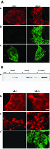
Figure 1.
Figure 1.
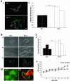
Cellular models. (A) Primary enterocytes from vil+/+ and vil-/- mice were cultured for 6 d. Epithelial islets were characterized using F-actin (red) (a) and villin (green) (b) double staining. Cytokeratin (green) labeling was used to check the epithelial origin of enterocytes islets (c). (B) Tightly controlled expression of villin by doxycycline (dox) treatment in MDCK cells. Cells grown in presence of the indicated doxycycline concentrations were separated into vil+/+ cells, grown in absence of the antibiotic, and vil-/- cells, grown in presence of doxycycline. Villin expression repression and induction was evaluated by Western blotting. This figure shows that villin induction is efficient when the cells are routinely grown in presence of 0.5 μg/μl doxyxycline (a). However, to avoid leaky expression during repression, the cells are shifted to 2 μg/μl doxycycline. Control of villin expression was also tested by immunofluorescence. Actin was labeled with phalloidin TRITC (b) and villin by using a mAb (c). Bar, 10 μm.
Figure 2.
Figure 2.
Villin enhances HGF-induced cell motility. (A) Primary enterocytes from vil+/+ and vil-/- mice were cultured for 6 d. Cells were stimulated with 10 IU/ml HGF to allow cell scattering. Individual cell trajectories were followed for 13 h after HGF stimulation (a) and analyzed using MetaMorph software to determine cell velocity data in micrometers per minute (b) (two independent experiments; *p < 0.05). (B) Vil-/- and vil+/+ MDCK cells before (a) and after9hofHGF(10 IU/ml) stimulation (b). c illustrates cell tracking after9hof treatment. (C) a, vil-/- and vil+/+ MDCK cell velocities upon HGF stimulation (three independent experiments; *p < 0.05). b, quantitation of lamellipodia extension as a function of time. The cell area was measured using MetaMorph software. (p = 0.011 at 140 min; p = 0.007 at 160 min). D. MDCK cells grown as small islets were stimulated with HGF (10 IU/ml) for 6 h. a, villin labeling in HGF-stimulated MDCK cells. b, villin colocalization with actin upon HGF stimulation. Cells were fixed and stained for villin (green) and actin (red). a and b, upon HGF stimulation, villin is redistributed to the leading edge of migrating cells and colocalizes with actin in this specialized region. Bar, 10 μm.
Figure 3.
Figure 3.
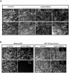
Villin induces F-actin reorganization in vitro. Rhodamine-labeled actin filaments were prepared as described in MATERIALS AND METHODS. They were allowed to attach to a nitrocellulose matrix on a glass coverslip mounted as a flow chamber during 15 min on ice. After washing with an antibleaching buffer, 12 μl of cell lysate (10 mg/ml) was perfused and the analysis was directly performed under fluorescent microscopy. (A) Enterocyte lysates. a and e, rhodamine-labeled F-actin filaments before lysate perfusion as internal controls. Note that in absence of any perfusion, the actin filaments correspond to disorganized actin network. B, 3 min after vil+/+ lysate addition. c, 5 min after EGTA treated vil+/+ lysate addition. f, 3 min after vil-/- lysate addition. g, 5 min after EGTA-treated vil-/- lysate addition. d, perfusion of vil-/- lysate followed by addition of 1 μg/μl recombinant villin during 5 min. h, actin filaments >1 h after vil-/- lysate perfusion. (B) MDCK cells lysates. a, c, e, and g, actin filaments before lysate perfusion. In absence of HGF, vil-/- (b) and vil+/+ (f) lysates. After 6 h of HGF stimulation, vil-/- (d) and vil+/+ (h) lysates. Bar, 10 μm.
Figure 4.
Figure 4.
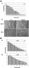
Villin expression enhances HGF-induced wound repair. (A) Wound-healing experiments performed on primary cultures of enterocytes. The wound has been performed using a thin needle on islets of enterocytes. a, measurement of the wound area over time. b, phase contrast image of enterocytes at T0 and 320 min after wound. (B) Wound-healing experiments on confluent monolayers of MDCK cells. The wound has been performed using a tip. a and b, measurement of the wound area over time in the absence of HGF (a) and in presence of HGF (10 UI/ml) (b). In both cases, wound repair was expressed as a percentage of the initial wound area.
Figure 5.
Figure 5.
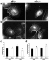
Villin-expressing cells present a decrease of the F-actin content and a higher level of barbed ends at the leading edge of MDCK cells upon HGF stimulation. Free barbed ends were visualized by rhodamine G-actin incorporation before (a) and after 6 h of HGF treatment (b). The amount of free barbed ends at the leading edge of the cells was evaluated by quantitation of the average fluorescence intensity using MetaMorph software (d). Eighteen vil-/- and 24 vil+/+ unstimulated cells were analyzed. Twentyseven vil-/- and 30 vil+/+ cells after 6 h of HGF treatment were analyzed (*p = 0.004). The F-actin content was evaluated by measuring the fluorescence intensity of TRITC-palloidin–labeled actin filaments in MDCK cells before and after 3 h of HGF treatment (c). The amount of total protein was the same in vil-/- and vil+/+ cells; n = 3 experiments.
Figure 6.
Figure 6.
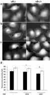
Actin dynamics of villin-expressing cells relies on a higher available pool of actin monomers than in repressed cells. The G-actin pool was visualized using fluorescent DNase I, which binds to actin monomers with a high affinity. Cells were either unstimulated (a) or stimulated with HGF (10 UI/ml) for 2 h (b) and 6 h (c). Arrows show maintenance of G-actin labeling in the leading edge of induced cells after 6 h of HGF treatment. Fluorescence intensity in the lamellipodia was quantified using MetaMorph software (d). Among the induced cells, 26 nonstimulated cells, 31 cells treated with HGF for 2 h, and 30 cells treated with HGF for 6 h were analyzed. Among the repressed cells, 31 nonstimulated cells, 36 cells treated with HGF for 2 h, and 30 cells treated with HGF for 6 h were analyzed. (p = 0.026 at 2 h; p = 0.001 at 6 h).
Figure 7.
Figure 7.
Tubulogenesis is enhanced in villin expressing MDCK cells. Immunofluorescence on collagen gels. a and a′, two confocal sections of E-cadherin labeled vil-/- tubules. b, double labeling of E cadherin and laminin. c and c′, two confocal sections of villin labeled vil+/+ tubules. d, double villin and laminin labeling. Laminin labeling is used to check for the differentiation status of the tubules. Bar, 40 μm.
Figure 8.
Figure 8.
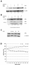
HGF stimulation induces villin association to the plasma membrane and increases its association to PLCg. (A) Villin redistribution in the membrane fraction upon HGF stimulation. This figure presents villin and E-cadherin blotting, shown as a control for equal loading. Cells were either unstimulated (lane 2), stimulated with HGF for 2 h (lane 3), treated with U73122 (10 μM) to inhibit PLCγ (lane 5), or double-treated with HGF and U73122 (lane 4). (B) Coimmunoprecipitation of villin and PLCγ in the membrane fraction. The first panel presents villin immunoprecipitation, blotted for villin. The second panel presents PLCγ immunoprecipitation blotted either for villin or for tyrosine phosphorylated proteins. The tyrosine-phosphorylated protein detected was at the molecular weight of villin. For all panels, the lanes are the following: lane 1, unstimulated cells; lane 2, after 2 h of HGF stimulation; lane 3, after U73122 (10 μM) treatment, lane 4, double treated with HGF and U73122; and lane 5, unstimulated cells but treated with pervanadate to induce protein phosphorylation. (C) Villin and α-tubulin blotting in total cell lysates before and after HGF treatment. (D) Lamellipod extension assays in presence of HGF in vil-/- and vil+/+ cells pretreated with U73122 (10 μM). Data are represented as the percentage of the initial cell area (T0).

Similar articles

Cited by

References

    1. Arpin, M., Pringault, E., Finidori, J., Garcia, A., Jeltsch, J.M., Vandekerckhove, J., and Louvard, D. (1988). Sequence of human villin: a large duplicated domain homologous with other actin-severing proteins and a unique small carboxy-terminal domain related to villin specificity. J. Cell Biol. 107, 1759-1766. - PMC - PubMed
    1. Athman, R., Louvard, D., and Robine, S. (2002a). The epithelial cell cytoskeleton and intracellular trafficking. III. How is villin involved in the actin cytoskeleton dynamics in intestinal cells? Am. J. Physiol. 283, G496-G502. - PubMed
    1. Athman, R., Niewohner, J., Louvard, D., and Robine, S. (2002b). Epithelial cells: establishment of primary cultures and immortalization. In: Molecular Cellular Microbiology, vol. 31, ed. P.S.a.A. Zychlinsky, New York: Academic Press, 93-113.
    1. Bhargava, M.M., Li, Y., Joseph, A., Jin, L., Rosen, E.M., and Goldberg, I.D. (1993). HGF-SF: effects on motility and morphology of normal and tumor cells. EXS 65, 341-349. - PubMed
    1. Boyer, B., Valles, A.M., and Thiery, J.P. (1996). Model systems of epitheliummesenchyme transitions. Acta Anat. 156, 227-239. - PubMed

Publication types

MeSH terms