. 2003 Oct;14(10):4221-9.
doi: 10.1091/mbc.e03-02-0111. Epub 2003 Aug 22.

DNA damage modulates nucleolar interaction of the Werner protein with the AAA ATPase p97/VCP

Affiliations

DNA damage modulates nucleolar interaction of the Werner protein with the AAA ATPase p97/VCP

Juneth Joaquin Partridge et al. Mol Biol Cell. 2003 Oct.

Abstract

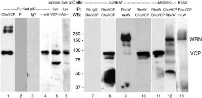
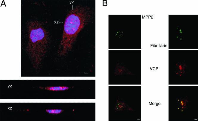
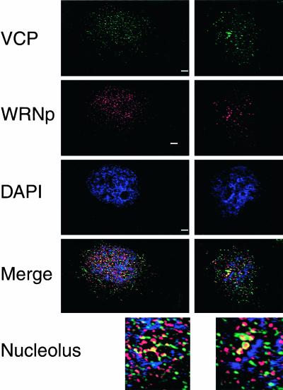
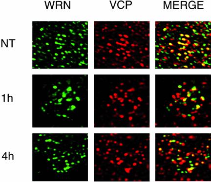
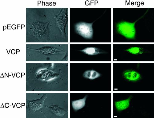
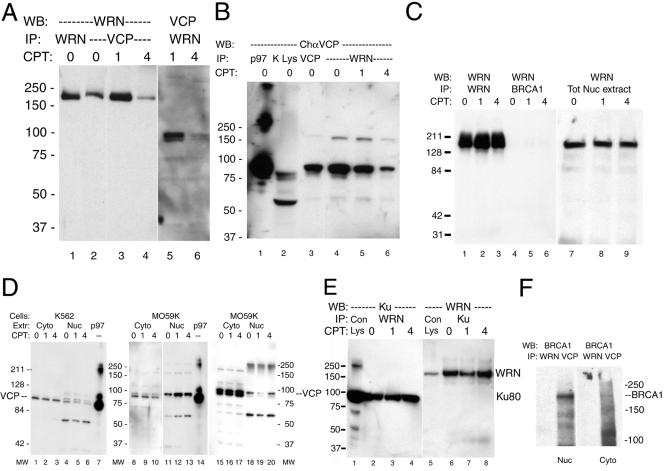
We report a novel nucleolar interaction between the AAA ATPase p97/VCP and the Werner protein (WRNp), a member of the RecQ helicase family. p97/VCP mediates several important cellular functions in eucaryotic cells, including membrane fusion of the endoplasmic reticulum and Golgi and ubiquitin-dependent protein degradation. Mutations in the WRN gene cause Werner syndrome, a genetic disorder characterized by premature onset of aging symptoms, a higher incidence of cancer, and a high susceptibility to DNA damage caused by topoisomerase inhibitors. We observed that both WRNp and valosin-containing protein (VCP) were present in the nucleoplasm and in nucleolar foci in mammalian cells and that WRNp and p97/VCP physically interacted in the nucleoli. Importantly, the nucleolar WRNp/VCP complex was dissociated by treatment with camptothecin, an inhibitor of topoisomerase I, whereas other WRNp-associated protein complexes, such as WRNp/Ku 80, were not dissociated by this drug. Because WRN syndrome cells are sensitive to topoisomerase inhibitors, these observations suggest that the VCP/WRNp interaction plays an important role in WRN biology. We propose a novel role for VCP in the DNA damage response pathway through modulation of WRNp availability.


Figures

Figure 4.
Figure 4.
VCP and WRNp reciprocally coimmunoprecipitate. Nuclear extracts were immunoprecipitated and immunoblotted as described in MATERIALS AND METHODS. Equal amounts of total protein were immunoprecipitated with 20 μl of rabbit anti-Werner helicase (RbαW) and either the chicken (1469) or rabbit (5860) anti-VCP polyclonal antibodies. Rabbit IgG was used as a negative control. Immunoprecipitated proteins (40 μl/lane) were electrophoresed on a 7.5% polyacrylamide gel and then immunoblotted to polyvinylidene difluoride membranes. After a 1 h incubation with primary antibodies and appropriate horseradish peroxidase-conjugated secondary antibodies, proteins were visualized by enhanced chemiluminescence. The results are presented as a composite image of Jurkat (lanes 7–10), MO59K (lanes 11 and 12) or K562 (lane 13) precipitates, or total cell lysates of MO56K (lane 5) or SW13 (lane 6) cells. Lanes 1–4 contain purified bovine liver VCP, 0.5 μg, immunoblotted with chicken anti-VCP 1469 (1:2000, lane 1), or anti-VCP mAb (lane 4). As a control, 2.0 μg of purified VCP was immunoblotted with 10 μg/ml preimmune chicken serum (preimmune 1:200, lane 2) or 10 μg/ml purified chicken IgY (IgY, lane 3). The expected position of VCP and WRNp are indicated on the right and molecular masses in kilodaltons are in the middle. In some cell lysates (example, lane 5), an ∼60-kDa protein, apparently a VCP fragment, is detected by anti-VCP antibody. VCP multimers are occasionally detected, for example, in purified VCP (lane 1).
Figure 1.
Figure 1.
VCP is present in the nucleus and nucleolus of mammalian cells. Cells were grown on glass coverslips overnight and then fixed in 3.7% formaldehyde and permeabilized with Triton X-100 as described in MATERIALS AND METHODS. (A) Confocal section of CV-1 cells stained with Rb 5860 anti-VCP (1:500) and visualized with Cy3-conjugated secondary antibody (red), and with the DNA stain DAPI (blue). xz and yz are confocal cross sections through the right-hand cell as marked. 630×; bar, 10 μm. (B) NCI-H226 cells were stained with chicken 1469 anti-VCP (1:200, red) and mouse anti-MPP2 (1:500, green) or mouse anti-fibrillarin (1:200, green). Images were merged to view colocalization (yellow). 400×; bar, 10 μm.
Figure 3.
Figure 3.
VCP and WRNp colocalize in the nucleolus. MO59K cells were processed for indirect immunofluorescence as described above and stained with anti-WRNp mAb (1:100, red), chicken 1469 anti-VCP (1:250, green), and the DNA stain DAPI (blue). By using Openlab deconvolution software, 0.1-μm Z-sections were obtained on an Axioplan 2 microscope (Carl Zeiss) and deconvolved. Merge is the merged image of the three fluorescence channels examined of the same deconvolved Z-section. Two representative cells from different experiments are shown; a nucleolar area of each cell was enlarged (3×, bottom). 630×; bar, 5 μM.
Figure 6.
Figure 6.
CPT treatment dissociates VCP and WRNp in the nucleolus. MO59K cells were treated with 10 μM CPT for 1 or 4 h or were NT and then processed for indirect immunofluorescence as described in Figure 3. WRN (green) and VCP (red) are the different fluorescent channels of the same 0.1-μm section examined. Merged is the result of coloring both channels and presenting them in the same image, with yellow indicating colocalization. NT, 630×; 1 and 4 h, 1000×.
Figure 2.
Figure 2.
The N-terminal domain of VCP is crucial for nuclear localization. The murine VCP gene was fused to the pEGFP vector as described in MATERIALS AND METHODS, and several domain-deleted fusion proteins were constructed as described in text. Human glioma MO59K cells were transfected using the cationic lipid method. After 48 h, live cells were examined under an Olympus IX70 and images captured and merged with IPLab. Right, merged image of GFP fluorescence (middle) with phase contrast (left). 400×, bar, 5 μm.
Figure 5.
Figure 5.
CPT treatment effects VCP and WRNp coimmunoprecipitates. MO59K cells were treated with 10 μM CPT for 1 or 4 h or were untreated controls (0 h). In all panels, the molecular mass in kilodaltons is indicated to the right or left. (A) Rabbit anti-WRNp and rabbit anti-VCP precipitates were treated as described in Figure 4 and Western blotted with mouse anti-WRNp (lanes 1–4) or chicken anti-VCP (lanes 5 and 6) as described. (B) A single immunoblot from the same experiment of VCP (lane 3) and WRNp (lanes 4–6) IPs probed with chicken anti-VCP. Lane 1, 0.5 μg of purified bovine liver VCP; lane 2, MO59K total cell lysate (40 μg). (C) Mouse anti-WRNp detects WRNp in rabbit anti-WRNp precipitates (lanes 1–3) and in total nuclear extract (lanes 7–9), but not in rabbit anti-BRCA1 IPs (lanes 4–6). Cells were treated with CPT and Western blotted as described in text. (D) Cells were treated as described above with CPT and extracts were immunoblotted with chicken 1469 anti-VCP as described in text. Three different experiments are shown, one performed with K562 extracts (lanes 1–6) and two with MO59K extracts (lanes 8–13 and 15–20). Lanes 7 and 14, 0.5 μg of purified bovine liver VCP. (E) WRNp was detected in rabbit anti-Ku 80 precipitates (lanes 6–8) and Ku-80 was detected in rabbit anti-WRNp precipitates (lanes 2–4) of nuclear extracts. Lane 1, 20 μl of SW13 total lysate; lane 5, 20 μl of MO59K total lysate. (F) BRCA1 detected in VCP, but not WRNp IPs. MO59K cells were separated into cytoplasmic and nuclear extracts as described in MATERIALS AND METHODS and precipitated with rabbit anti-WRNp or Rb 5860 anti-VCP and probed for BRCA1.

References

    1. Abmayr, S.M., and Workman, J.L. (2001). Unit 12.1 Preparation of nuclear and cytoplasmic extracts from mammalian cells. In: Current Protocols in Molecular Biology Online, ed. F.M. Ausubel, New York: John Wiley & Sons. - PubMed
    1. Acharya, U., Jacobs, R., Peters, J.M., Watson, N., Farquhar, M.G., and Malhotra, V. (1995). The formation of Golgi stacks from vesiculated Golgi membranes requires two distinct fusion events. Cell 82, 895–904. - PubMed
    1. Andersen, J.S., Lyon, C.E., Fox, A.H., Leung, A.K., Lam, Y.W., Steen, H., Mann, M., and Lamond, A.I. (2002). Directed proteomic analysis of the human nucleolus. Curr. Biol. 12, 1–11. - PubMed
    1. Blander, G., Kipnis, J., Leal, J.F., Yu, C.E., Schellenberg, G.D., and Oren, M. (1999). Physical and functional interaction between p53 and the Werner's syndrome protein. J. Biol. Chem. 274, 29463–29469. - PubMed
    1. Blander, G., Zalle, N., Leal, J.F., Bar-Or, R.L., Yu, C.E., and Oren, M. (2000). The Werner syndrome protein contributes to induction of p53 by DNA damage. FASEB J. 14, 2138–2140. - PubMed

Publication types

MeSH terms