. 2003 Oct;14(10):4140-54.
doi: 10.1091/mbc.e03-03-0167. Epub 2003 Aug 22.

Maximal polar growth potential depends on the polarisome component AgSpa2 in the filamentous fungus Ashbya gossypii

Affiliations

Maximal polar growth potential depends on the polarisome component AgSpa2 in the filamentous fungus Ashbya gossypii

Philipp Knechtle et al. Mol Biol Cell. 2003 Oct.

Abstract

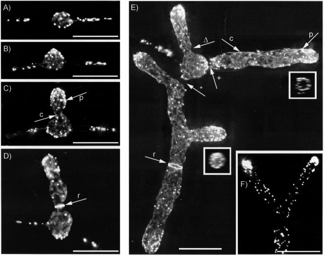
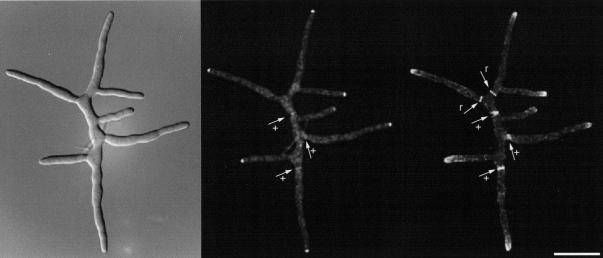
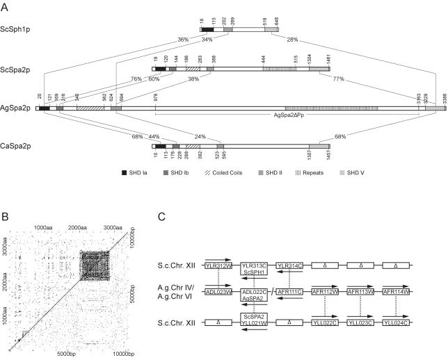
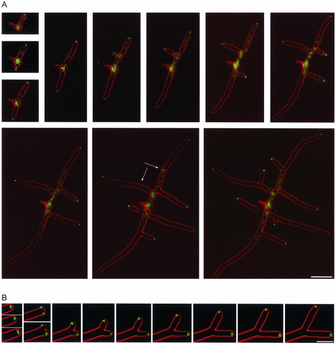
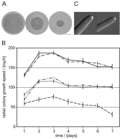
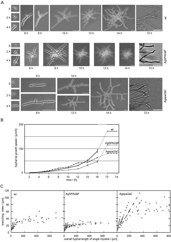
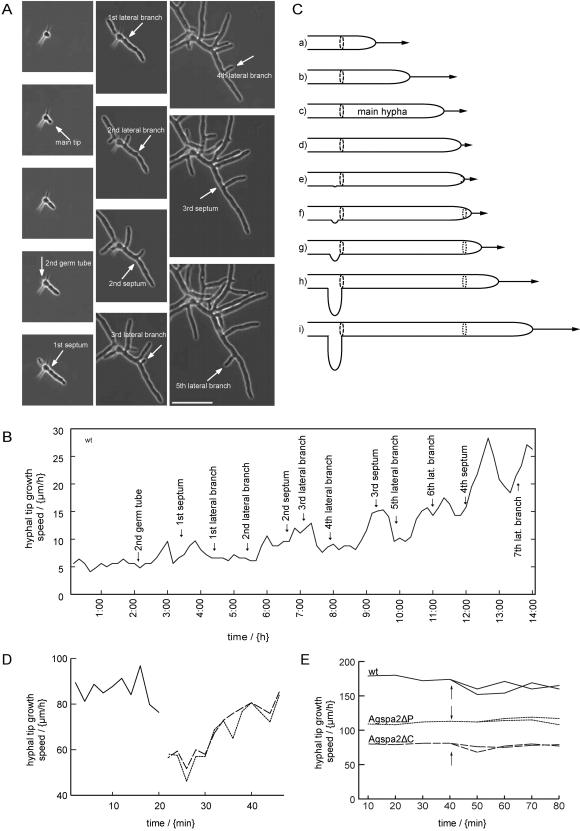
We used actin staining and videomicroscopy to analyze the development from a spore to a young mycelium in the filamentous ascomycete Ashbya gossypii. The development starts with an initial isotropic growth phase followed by the emergence of germ tubes. The initial tip growth speed of 6-10 microm/h increases during early stages of development. This increase is transiently interrupted in response to the establishment of lateral branches or septa. The hyphal tip growth speed finally reaches a maximum of up to 200 micro/h, and the tips of these mature hyphae have the ability to split into two equally fast-growing hyphae. A search for A. gossypii homologs of polarisome components of the yeast Saccharomyces cerevisiae revealed a remarkable size difference between Spa2p of both organisms, with AgSpa2p being double as long as ScSpa2p due to an extended internal domain. AgSpa2 colocalizes with sites of polarized actin. Using time-lapse videomicroscopy, we show that AgSpa2p-GFP polarization is established at sites of branch initiation and then permanently maintained at hyphal tips. Polarization at sites of septation is transient. During apical branching the existing AgSpa2p-GFP polarization is symmetrically divided. To investigate the function of AgSpa2p, we generated two AgSPA2 mutants, a partial deletion of the internal domain alone, and a complete deletion. The mutations had an impact on the maximal hyphal tip growth speed, on the hyphal diameter, and on the branching pattern. We suggest that AgSpa2p is required for the determination of the area of growth at the hyphal tip and that the extended internal domain plays an important role in this process.


Figures

Figure 1.
Figure 1.
A. gossypii rhodamine-phalloidin stainings during development from a spore to a mature mycelium. See text for description. The rectangles in Figure 1E show a horizontal view on the top of the respective tips. The most apical part of the tip is free of actin patches and forms a “hole” (see also the animated 3D reconstruction in Movie 1). Fluorescence can also be observed in the needle-shaped spores. The origin and importance of these structures is elusive. Bar, 10 μm. p, actin patch; c, actin cable; Δ, neck before septation; r, actin ring, a prerequisite of septum formation; +, actin patches on both sides of a growing septum; and *, completed septum lacking actin.
Figure 4.
Figure 4.
AgSpa2p-GFP/rhodamine-phalloidin double staining. Left, DIC; middle, AgSpa2p-GFP; and right, rhodamine-phalloidin. r, actin rings during early stages of septation and +, actin patches and AgSpa2p-GFP, respectively, at sites of septation during later developmental stages. See also Figure 1. Bar, 20 μm.
Figure 2.
Figure 2.
Analysis of AgSpa2p and its coding region. (A) Alignment of AgSpa2p, ScSpa2p, CaSpa2p, and ScSph1p. Corresponding domains show the same hatching. The domain homologies between AgSpa2p and ScSpa2p, CaSpa2p and ScSph1p, respectively, are given in percent identities. The position of the individual domains is marked in aa. The deleted region in AgSPA2ΔP is marked. In S. cerevisiae, ScSte11p, ScMkk1p, and ScKkk2p interact with SHD Ia (Sheu et al., 1998), ScPea2 with SHD II (Valtz and Herskowitz, 1996), and ScBni1p with SHD V (Fujiwara et al., 1998). (B) Comparison of repetitive regions in AgSpa2p and the AgSPA2 ORF. Left of the diagonal line shows a comparison of the AgSpa2p versus itself and right of the diagonal a comparison of the AgSPA2 ORF versus itself. (C) Syntenic analysis of the AgSPA2 gene. Each gene is represented as a rectangle and its orientation is indicated with an arrow.
Figure 3.
Figure 3.
Temporal organization of AgSpa2p-GFP during A. gossypii development. The GFP signal is indicated in green, the phase contrast in red. (A) Development from a spore to a young mycelium. The time elapsed between two frames is 1 h. Localization of AgSpa2p-GFP to the hyphal cortex before lateral branch emergence is indicated with arrows. The permanent localization pattern at hyphal tips was confirmed in two additional time-lapse acquisitions. As proven in still pictures, the cytoplasmic fluorescence observed toward the image center is an artifact induced by an uneven illumination of the sample. Refer also to supplemental Movie 2. Bar, 20 μm. (B) Apical branching after 20 h of development. The time elapsed between two frames is 2 min. Between frame 3 and 9 the GFP signal seems not to localize exactly to the hyphal tip. This is an artifact induced by different focal planes of the bright-field and the fluorescence image. This is also the reason for the weak GFP signal for the lower tip in frame 6. The localization pattern of AgSpa2p-GFP during apical branching was confirmed in one additional time lapse acquisition. Refer also to supplemental Movie 3. Bar, 10 μm.
Figure 5.
Figure 5.
Radial colony growth phenotype in AgSPA2ΔP and Agspa2ΔC strains. (A) Left, wild-type; middle, AgSPA2ΔP; and right, Agspa2ΔC. All mycelia were grown for 6 d at 30°C. Bar, 2 cm. (B) Radial growth speed of wild type, AgSPA2ΔP, and Agspa2ΔC strains. The x-axis represents the time in days and the y-axis the radial growth speed in micrometers per hour (error bar, SEM). Wild type (——), AgSPA2-GFP (– - –), AgSPA2ΔP (- -), AgSPA2ΔP-GFP (– - - –), and Agspa2Δ (– – –). The value measured after the 1st d was the difference between the inocula (1 mm in diameter) and the radial growth distance after 1 d divided by 24 h. The inocula were taken from plates that had already been growing for 3 d and should have reached the maximal radial growth speed. We suppose therefore that the value for day 1 (especially for the strains carrying the full-length and partially deleted alleles) is too low. (C) Localization of AgSpa2p-GFP (left) and AgSpa2ΔPp-GFP (right) to hyphal tips. DIC in gray and GFP in white.
Figure 6.
Figure 6.
Quantification of development from spores to mature mycelia in wild type, AgSPA2ΔP, and Agspa2ΔC. (A) Morphological analysis. The time elapsed between two frames is 2 h, whereas the last frame corresponds to the morphological stage after 72 h. Bar, 50 μm. (B) Hyphal tip growth speed. The x-axis represents the time in hours and the y-axis the hyphal tip growth speed in micrometers per hour. Wild type (——), AgSPA2ΔP (- -), and Agspa2ΔC(–––) (error bar, SEM; n > 20 for each point). (C) Branching index. The x-axis represents the total hyphal length of single mycelia and the y-axis the total hyphal length of single mycelia divided by the number of tips.
Figure 7.
Figure 7.
Behavior of the hyphal tip growth speed during the development of a spore to a mature mycelium. (A) Time-lapse acquisition of wild type at 2-min intervals. The time elapsed between the frames shown is 1 h. We followed the development of the first emerging germ tube (main tip). Sites of septation and lateral branch emergence are marked by arrows. We show the emergence of five lateral branches of nine observed and three septation events of four observed. Bar, 50 μm. Refer also to supplemental Movie 4. (B) Hyphal tip growth speed of wild type at medium resolution. The basis for this graph is the supplemental Movie 4, representative frames are shown in A. The x-axis represents the elapsed time in min and the y-axis the hyphal tip growth speed in micrometers per hour. Labeled arrows mark the initiation of septa and branches. Lateral branches show up 10–20 min after a hyphal tip growth speed decrease, whereas septa seem to occur slightly later. The reason for this might be that the beginning of a septum formation cannot be seen in phase contrast microscopy. We marked the emergence of seven lateral branches and four septations. This pattern was confirmed in two additional time lapse acquisitions. (C) Presumptive landmarks for lateral branching and septation in A. gossypii. The illustration shows the establishment of a lateral branch at a presumptive landmark (dashed ring) during development of a young A. gossypii mycelium. The outline of the hypha is indicated, and the changes in hyphal tip growth speed are represented by arrows of different lengths. During early development, the hyphal tip growth speed increases (a and b). Before lateral branch formation, the hyphal tip growth speed transiently decreases (c and d) and then accelerates again (e–i). The lateral branch emerges concomitant with the reacceleration of the main hypha or with a slight delay (e). During or after the transient decrease (c and d), a landmark is established at the tip (e, dotted line) that remains at the hyphal cortex (f–i, dotted ring) and marks a future branching and/or septation site. The timing for the establishment of new lateral branches and septa, respectively, and the decision whether to form a lateral branch or a septa are so far unclear. (D) Hyphal tip growth speed during apical branching in early development (16–20 h). This graph is based on a quantitative analysis of supplemental Movie 3, representative frames of which are shown in Figure 3B. The x-axis represents the elapsed time in minutes and the y-axis the hyphal tip growth speed in micrometers per hour. Apical branching occurs after 22 min with a concomitant decrease in tip growth speed of the two new hyphae. The solid line shows the hyphal tip growth speed before apical branching and the dashed and dotted lines the hyphal tip growth speeds after apical branching. This decrease and reacceleration was confirmed in one additional time lapse acquisition. (E) Lack of significant decrease in tip growth speed during apical branching in mature mycelium (3 d). Mature hyphae were followed by time lapse acquisitions in wild type, AgSPA2ΔP, and Agspa2ΔC strains (Figure 6A). The initiation of an apical branch is indicated with arrows. The hyphal tip growth speeds of both tips resulting from the apical branch were determined. Wild type (——), AgSPA2ΔP (- -), and Agspa2ΔC (– – –). Five independent apical branching events were observed for each strain giving similar results.

Similar articles

Cited by

References

    1. Altmann-Johl, R., and Philippsen, P. (1996). AgTHR4, a new selection marker for transformation of the filamentous fungus Ashbya gossypii, maps in a four-gene cluster that is conserved between A. gossypii and Saccharomyces cerevisiae. Mol. Gen. Genet. 250, 69–80. - PubMed
    1. Amberg, D.C. (1998). Three-dimensional imaging of the yeast actin cytoskeleton through the budding cell cycle. Mol. Biol. Cell 9, 3259–3262. - PMC - PubMed
    1. Amberg, D.C., Zahner, J.E., Mulholland, J.W., Pringle, J.R., and Botstein, D. (1997). Aip3p/Bud6p, a yeast actin-interacting protein that is involved in morphogenesis and the selection of bipolar budding sites. Mol. Biol. Cell 8, 729–753. - PMC - PubMed
    1. Arkowitz, R.A., and Lowe, N. (1997). A small conserved domain in the yeast Spa2p is necessary and sufficient for its polarized localization. J. Cell Biol. 138, 17–36. - PMC - PubMed
    1. Ayad-Durieux, Y., Knechtle, P., Goff, S., Dietrich, F., and Philippsen, P. (2000). A PAK-like protein kinase is required for maturation of young hyphae and septation in the filamentous ascomycete Ashbya gossypii. J. Cell Sci. 113, 4563–4575. - PubMed

Publication types

LinkOut - more resources