. 2003 Dec;14(12):5104-15.
doi: 10.1091/mbc.e03-04-0237. Epub 2003 Aug 22.

Caenorhabditis elegans nucleoporins Nup93 and Nup205 determine the limit of nuclear pore complex size exclusion in vivo

Affiliations

Caenorhabditis elegans nucleoporins Nup93 and Nup205 determine the limit of nuclear pore complex size exclusion in vivo

Vincent Galy et al. Mol Biol Cell. 2003 Dec.

Abstract

Nuclear pore complexes (NPCs) span the nuclear envelope and mediate communication between the nucleus and the cytoplasm. To obtain insight into the structure and function of NPCs of multicellular organisms, we have initiated an extensive analysis of Caenorhabditis elegans nucleoporins. Of 20 assigned C. elegans nucleoporin genes, 17 were found to be essential for embryonic development either alone or in combination. In several cases, depletion of nucleoporins by RNAi caused severe defects in nuclear appearance. More specifically, the C. elegans homologs of vertebrate Nup93 and Nup205 were each found to be required for normal NPC distribution in the nuclear envelope in vivo. Depletion of Nup93 or Nup205 caused a failure in nuclear exclusion of nonnuclear macromolecules of approximately 70 kDa without preventing active nuclear protein import or the assembly of the nuclear envelope. The defects in NPC exclusion were accompanied by abnormal chromatin condensation and early embryonic arrest. Thus, the contribution to NPC structure of Nup93 and Nup205 is essential for establishment of normal NPC function and for cell viability.


Figures
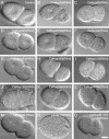
Figure 1.
Figure 1.
Depletion of nucleoporins causes nuclear morphology alterations in early embryos. Still images from time-lapse DIC microscopy are shown of a wild-type two-cell embryo (A), or of two-cell embryos depleted of CeNup54/NPP-1 (B), CeNup85/NPP-2 (C), CeNup205/NPP-3 (D), CeNup45/58/NPP-4 (E), CeNup62/NPP-11 (F), CeNup93/NPP-13 (G), CeNup160/NPP-6 (H), CeNup153/NPP-7 (I), CeNup155/NPP-8 (J), CeNup358/NPP-9 (K), CeNup98/96/NPP-10N/C (L), CeNup35/NPP-19 (M), CeSec13/NPP-20 (N), or triple depleted of CeNup85/NPP-2, CeNup107/NPP-5, and CeNup133/NPP-15 (O). Embryos are oriented with anterior on the left and similar time points relative to cytokinesis are displayed for all embryos. Bar, 10 μm.
Figure 2.
Figure 2.
Combinatorial RNAi reveals essential combinations of nucleoporins in the Nup107/Nup160 complex. The effect of RNAi on embryonic lethality was measured as described in MATERIALS AND METHODS. Embryonic lethality was measured upon RNAi against CeMAN1/LEM-2, CeNup107/NPP-5, CeNup133/NPP-15, CeNup107+CeNup133, CeNup214/NPP-14, or CeRAE1/NPP-17 alone (black bars, values from Table 1) or in combination with depletion of CeNup85/NPP-2 (hatched bars). CeNup85/NPP-2 alone resulted in 37% lethality, whereas targeting CeNup85/NPP-2 together with CeNup107/NPP-5, CeNup133/NPP-15, CeNup214/NPP-14, or CeRAE1/NPP-17 caused a significant increase in lethality. Triple RNAi against CeNup85/NPP-2, CeNup107/NPP-5, and CeNup133/NPP-15 similarly enhanced the lethality compared with CeNup85/NPP-2(RNAi) alone or CeNup107/NPP-5(RNAi) together with CeNup133/NPP-15(RNAi). Error bars indicate standard deviations.
Figure 3.
Figure 3.
Depletion of CeNup205 or CeNup93 delays cell division and induces abnormal peripheral chromatin condensation. (A) Embryos expressing YFP-lamin from control worms or worms depleted of CeNup205/NPP-3 or CeNup93/NPP-13 were observed by time-lapse confocal microscopy. Still images of two-cell embryos are shown with indication of time (minutes:seconds) relative to anaphase onset. Anterior of the embryos is on the left. In all cases, lamin accumulated at the nuclear periphery and in the nucleoplasm. (B) Maximum diameter of P1 nuclei was measured. Shown is the average of control, CeNup205(RNAi) or CeNup93(RNAi) embryos (n = 3 for each). (C) Timing of cell division was measured from P0 anaphase onset to AB anaphase onset (dark gray) and from AB anaphase onset to P1 anaphase onset (light gray). Shown is the average of control, CeNup205(RNAi) or CeNup93(RNAi) embryos (n = 3 for each). (D) Embryos expressing GFP-histone H2B from control worms or from worms depleted of CeNup205/NPP-3 or CeNup93/NPP-13 were observed by confocal microscopy. Shown are projections from z-scan acquisitions (z step = 0.5 μm) of representative CeNup205(RNAi) or CeNup93(RNAi) embryos collected 16 h after oviposition or a wild type embryo with a comparable cell number. Note the heterogeneous distribution and the abnormal peripheral condensation of chromatin in CeNup205(RNAi) and CeNup93 (RNAi) embryos. (E) Embryos expressing GFP-β-tubulin and GFP-histone H2B from either control worms or worms depleted of CeNup205/NPP-3 or CeNup93/NPP-13 were observed by time-lapse confocal microscopy. Still images are shown with indication of time (minutes:seconds) relative to anaphase onset. Note the abnormal peripheral condensation of chromatin in P1 of 2-cell CeNup205(RNAi) and CeNup93(RNAi) embryos (indicated by arrows). Bars, 10 μm.
Figure 4.
Figure 4.
Depletion of CeNup205 or CeNup93 induces abnormal NPC distribution in the NE. (A) Embryos from control worms or worms depleted of CeNup205/NPP-3 or CeNup93/NPP-13 were fixed and analyzed with the anti-nucleoporin mAb414. Projections from z-scan acquisitions by using confocal microscopy (z step = 0.5 μm) at low (first row; bar, 10 μm) or higher (second row; bar, 1 μm) magnification are shown. NPCs are clustered in the NE of CeNup205/NPP-3- or CeNup93/NPP-13–depleted embryos. (B) Embryos expressing GFP-MAN1 from control worms or worms depleted of CeNup205/NPP-3 or CeNup93/NPP-13 were fixed and observed by confocal microscopy. Single confocal sections are shown. Note that the GFP-MAN1 distribution was uniform in the NEs of all embryos.
Figure 5.
Figure 5.
Depletion of CeNup205 or CeNup93 increases the size range of macromolecules that can diffuse freely across the NPC. (A) Young embryos expressing GFP-β-tubulin from control worms or worms depleted of CeNup205/NPP-3 or CeNup93/NPP-13 were observed by time-lapse confocal microscopy. Still images are shown with indication of time (minutes:seconds) relative to anaphase onset. Anterior of the embryos is on the left. Soluble GFP-β-tubulin was not excluded form the nuclear space in CeNup205(RNAi) or CeNup93(RNAi) embryos. Pronuclei in CeNup205(RNAi) embryos also failed to exclude GFP-β-tubulin. (B) Soluble GFP-β-tubulin was not excluded from the nuclear space in older CeNup205(RNAi) or CeNup93(RNAi) embryos. (C and D) Gonads of control worms or worms depleted of CeNup205 were injected with a mix of fluorescent dextrans of 10 kDa (left) and 70 kDa (right) (C) or a mix of fluorescent dextrans of 10 kDa (left) and 160 kDa (right) (D). Dextran distribution in embryos was analyzed by confocal microscopy. Single confocal images are shown. The 10-kDa dextran diffused freely into all nuclei, whereas the 70-kDa dextran was only excluded from nuclei of control embryos and not from nuclei of CeNup205(RNAi) embryos (C). The 160-kDa dextran is excluded from the nuclei of both control embryos and CeNup205(RNAi) embryos. Bar, 10 μm.

Similar articles

Cited by

References

    1. Antonny, B., and Schekman, R. (2001). ER export: public transportation by the COPII coach. Curr. Opin. Cell Biol. 13, 438-443. - PubMed
    1. Askjaer, P., Galy, V., Hannak, E., and Mattaj, I.W. (2002). Ran GTPase cycle and importins alpha and beta are essential for spindle formation and nuclear envelope assembly in living Caenorhabditis elegans embryos. Mol. Biol. Cell 13, 4355-4370. - PMC - PubMed
    1. Bagley, S., Goldberg, M.W., Cronshaw, J.M., Rutherford, S., and Allen, T.D. (2000). The nuclear pore complex. J. Cell Sci. 113, 3885-3886. - PubMed
    1. Belgareh, N., et al. (2001). An evolutionarily conserved NPC subcomplex, which redistributes in part to kinetochores in mammalian cells. J. Cell Biol. 154, 1147-1160. - PMC - PubMed
    1. Boehmer, T., Enninga, J., Dales, S., Blobel, G., and Zhong, H. (2003). Depletion of a single nucleoporin, Nup107, prevents the assembly of a subset of nucleoporins into the nuclear pore complex. Proc. Natl. Acad. Sci. USA 100, 981-985. - PMC - PubMed

Publication types

MeSH terms

LinkOut - more resources