. 2003 Nov;14(11):4397-413.
doi: 10.1091/mbc.e03-05-0315. Epub 2003 Aug 22.

Impairing actin filament or syndapin functions promotes accumulation of clathrin-coated vesicles at the apical plasma membrane of acinar epithelial cells

Affiliations

Impairing actin filament or syndapin functions promotes accumulation of clathrin-coated vesicles at the apical plasma membrane of acinar epithelial cells

Silvia R Da Costa et al. Mol Biol Cell. 2003 Nov.

Abstract

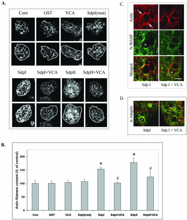
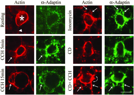
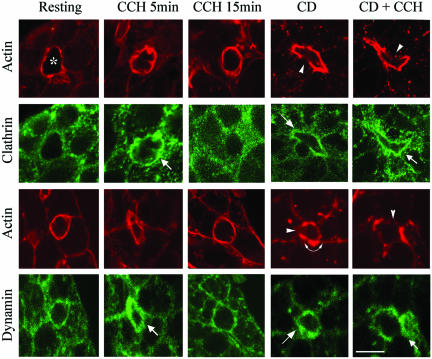
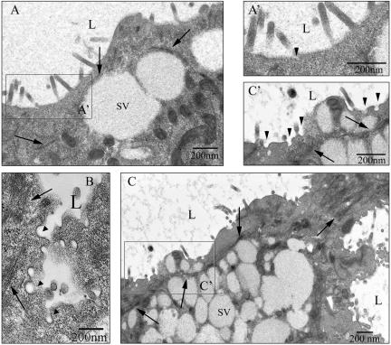
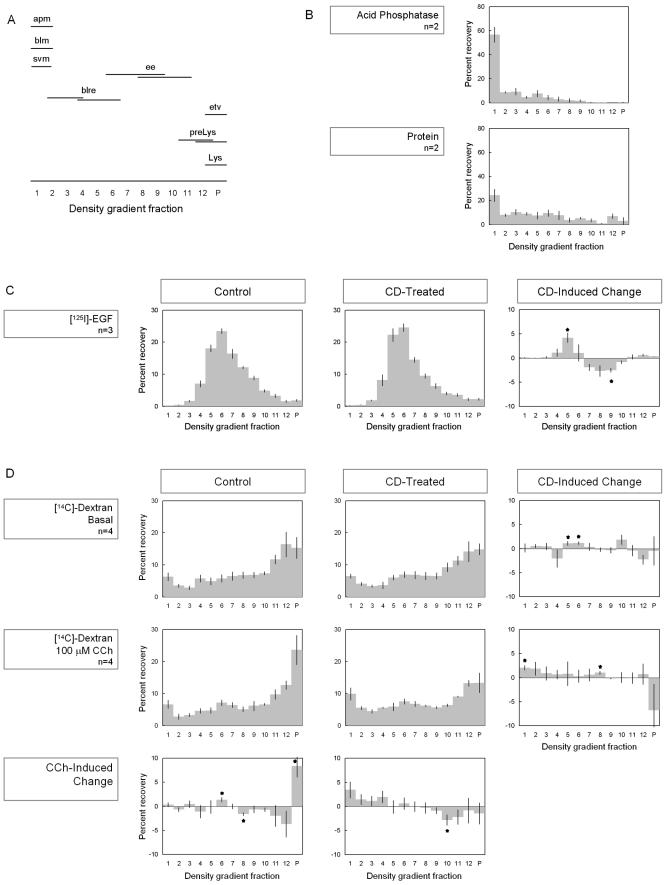
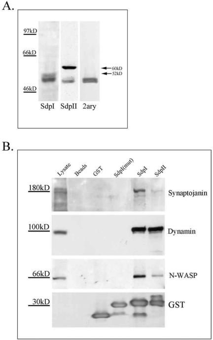
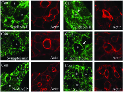
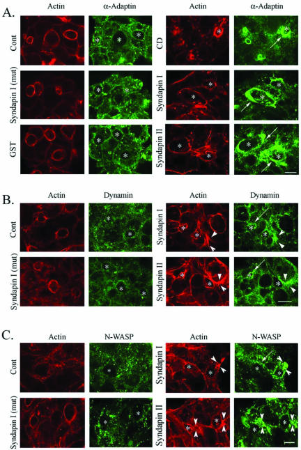
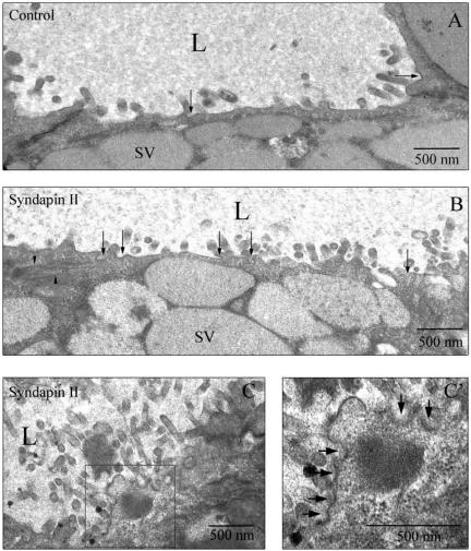
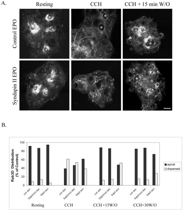
In this article, we investigate the contributions of actin filaments and accessory proteins to apical clathrin-mediated endocytosis in primary rabbit lacrimal acini. Confocal fluorescence and electron microscopy revealed that cytochalasin D promoted apical accumulation of clathrin, alpha-adaptin, dynamin, and F-actin and increased the amounts of coated pits and vesicles at the apical plasma membrane. Sorbitol density gradient analysis of membrane compartments showed that cytochalasin D increased [14C]dextran association with apical membranes from stimulated acini, consistent with functional inhibition of apical endocytosis. Recombinant syndapin SH3 domains interacted with lacrimal acinar dynamin, neuronal Wiskott-Aldrich Syndrome protein (N-WASP), and synaptojanin; their introduction by electroporation elicited remarkable accumulation of clathrin, accessory proteins, and coated pits at the apical plasma membrane. These SH3 domains also significantly (p </= 0.05) increased F-actin, with substantial colocalization of dynamin and N-WASP with the additional filaments. Coelectroporation with the VCA domain of N-WASP blocked the increase in F-actin and reversed the morphological changes indicative of impaired apical endocytosis. We suggest that transient modulation of actin polymerization by syndapins through activation of the Arp2/3 complex via N-WASP coordinates dynamin-mediated vesicle fission at the apical plasma membrane of acinar epithelia. Trapping of assembled F-actin intermediates during this process by cytochalasin D or syndapin SH3 domains impairs endocytosis.


Figures

Figure 9.
Figure 9.
Analysis of F-actin content in acini electroporated with GST fusion proteins. (A) Representative serial sections of acini electroporated without (control) or with GST fusion proteins as indicated and then fixed and labeled with rhodamine-phalloidin. Sections depicted for each treatment were acquired at ≅1-μm intervals and are representative of the serial sectioning used for quantitative analysis (see MATERIALS AND METHODS). (B) Quantitation of the intensity of actin filament labeling under each condition was obtained using MetaMorph Quantitation Software as described in MATERIALS AND METHODS (10 images/acinus, four acini, and 160 images total/treatment, n = 3–6 separate preparations; *p ≤ 0.05, Bar, ∼10 μm). Distribution of N-WASP (green) in parallel with actin filaments (red) (C) and distribution of α-adaptin (green) in parallel with actin filaments (red) (D) as detected by confocal fluorescence microscopy in resting acini electroporated with syndapin I SH3 domains without (left) or with (right) the GST-VCA fusion protein. Apical/lumenal regions are identified in each panel by *. Arrows depict the accumulation of N-WASP with additional F-actin structures formed in acini electroporated with the syndapin I SH3 domain. Bars, ∼10 μm.
Figure 1.
Figure 1.
CD promotes accumulation of α-adaptin at the APM of lacrimal acini. The intracellular distribution of α-adaptin (green) in control and stimulated acini with and without CD was investigated using anti-α-adaptin primary and FITC-conjugated secondary antibodies and detected by confocal fluorescence microscopy. F-Actin organization (red) was probed in parallel in each sample using rhodamine-phalloidin. Top left, representative lumenal region (*) surrounded by APM underlaid with abundant apical actin filaments and fainter labeling indicative of basolateral actin (large arrow). Treatments included 100 μM CCH for either 5 or 15 min without or with CD (5 μM, 60 min) or 1 μM ionomycin for 5 min. Small arrows in paired images denote sites of accumulation of actin-coated SVs and α-adaptin. Arrowhead indicates basolateral actin and curved arrow denotes regions of apparent actin accumulation. Bar, ∼10 μm.
Figure 2.
Figure 2.
CD promotes accumulation of clathrin and dynamin at the APM of lacrimal acini. The intracellular distributions of clathrin or dynamin (green) in control and CCH-stimulated acini with and without CD were investigated using appropriate primary and FITC-conjugated secondary antibodies and detected by confocal fluorescence microscopy. F-Actin organization (red) was probed in parallel in each sample by using rhodamine-phalloidin. Apical/lumenal regions are identified by the enrichment of F-actin, denoted by (*) in the top left image. Treatments included 100 μM CCH for either 5 or 15 min without or with CD (5 μM, 60 min). Small arrows indicate apparent accumulation of clathrin or dynamin beneath apparent actin-coated SVs, arrowheads denote regions of apparent actin fragmentation associated with CD, and curved arrow denotes regions of apparent actin accumulation. Bar, ∼10 μm.
Figure 3.
Figure 3.
CD increases coated pits at the APM of lacrimal acini while also bundling some apical actin filaments. Resting acini (A) and acini exposed to CD (5 μM, 60 min) with (B) or without (C) CCH stimulation (100 μM, 15 min). Acini were cultured on Matrigel rafts and processed for EM as described in MATERIALS AND METHODS. Lumena (L) are enriched in mature SVs and filamentous structures (arrows), which are likely to be actin filaments. Apparent coated pits and coated vesicles (arrowheads) are detected in trace amounts in resting acini (A, A′) acini but are increased dramatically in CD-treated acini (C and C′). Exposure of CD-treated acini to CCH also resulted in accumulation of coated pits and vesicles (arrowheads) in regions apparently undergoing exocytosis and enriched in actin filaments (arrows) (B).
Figure 4
Figure 4
CD increases association of [14C]dextran with APM fractions. (A) Working model for isolation of acinar plasma membrane and endocytic membrane compartments over sorbitol density gradients (apm, apical plasma membrane; blm, basolateral membrane; svm, SV membrane; blre, basolateral recycling endosome; ee, early endosome; etv, endocytic transport vesicle; preLys, prelysosome; Lys, lysosome). (B) Lacrimal glands were homogenized and subjected to Mg2+ precipitation of APM, and the resulting membrane sample was processed by isopycnic centrifugation on sorbitol density gradients and fractions analyzed for protein and acid phosphatase activity. (C) 125I-EGF content of membrane fractions isolated by isopycnic centrifugation on sorbitol density gradients from resting acini or acini exposed to CD (5 μM, 60 min). The right column shows the CD-induced change. (D) [14C]Dextran content of membrane fractions isolated by isopycnic centrifugation on sorbitol density gradients from resting and CCH-stimulated acini (100 μm, 15 min) with and without CD (5 μM, 60 min). The right column shows the CD-induced change, whereas the bottom row shows the CCH-induced change. n = 2–4 preparations as indicated; error bars represent SEM; *p ≤ 0.05.
Figure 5.
Figure 5.
Syndapin I and II abundance and binding partners in acini. (A) SDS-PAGE of lacrimal gland acinar lysates (150 μg of protein/lane) and subsequent Western blotting with appropriate primary and secondary antibodies as indicated revealed bands at 5 and 60 kDa consistent with the presence of syndapins I and II, respectively, in lacrimal acini. The goat-anti rabbit secondary antibody (2ary) used for these Western blots reacts with a 50-kDa protein present in rabbit acinar lysates. (B) Affinity purifications of proteins interacting with the SH3 domains of syndapins I and II, the mutated SH3 domain of syndapin I, GST, and glutathione-agarose beads alone. Equal amounts of fusion proteins (75 μg of fusion protein/sample) and lacrimal acinar lysate (≅4.3 mg protein/sample) were used for each pull-down sample. Beads were resuspended in 150 μl of sample buffer, which also solubilized the GST fusion proteins associated with the beads, and 35 μl of sample was loaded in each case. Starting material (91 μg) was loaded for comparison. Western blots were probed with appropriate primary antibodies against synaptojanin, dynamin, N-WASP, and GST.
Figure 6.
Figure 6.
Distributions of syndapins I and II, synaptojanin-I, and N-WASP in acini. The distributions of syndapins I and II, synaptojanin, and N-WASP (green) were investigated using appropriate primary and fluorophoreconjugated secondary antibodies and detected by confocal fluorescence microscopy. F-actin organization (red, matched image) was probed in parallel in each sample by using rhodamine-phalloidin. The distribution of each of these proteins is shown in resting acini (Con), whereas the distribution of syndapin II in CD-treated acini (5 μM, 60 min) and the distribution of synaptojanin in CCH-treated acini (100 μM, 5 min) are also indicated. Enrichment of these proteins at the APM is indicated by arrows. Apical/lumenal regions are identified in each panel by *. Bar, ∼10 μm.
Figure 7.
Figure 7.
Syndapin SH3 domains elicit specific accumulation of α-adaptin and dynamin at the APM of acini. The distributions of α-adaptin (green, A), dynamin (green, B) and N-WASP (green, C) in parallel with actin filaments (red, all panels) in resting acini electroporated with GST fusion proteins were probed using appropriate primary and fluorophore-conjugated secondary antibodies and rhodamine-phalloidin. The apical accumulation of α-adaptin elicited by CD treatment (5 μM, 60 min) is also shown for comparison. Apical/lumenal regions are identified by *. Arrows depict the accumulation of α-adaptin, dynamin, or N-WASP, respectively, at the APM associated with CD treatment or introduction of syndapin SH3 domains. Arrowheads depict the accumulation of dynamin or N-WASP in parallel with additional F-actin structures elicited by electroporation with syndapin SH3 domains. Bar, ∼10 μm.
Figure 8.
Figure 8.
EM images of coated pits and vesicles at the APM of resting acini electroporated with GST fusion proteins. (A) APM-enriched in microvilli facing a lumen (L) in an acinus electroporated without fusion proteins. Occasional clathrin-coated pits or vesicles (arrows) are seen at the APM. (B) Introduction of the syndapin II SH3 domain increased the number of clathrin-coated pits or vesicles (arrows) detected at the APM as well as actin bundles (arrowheads) (C) Acini electroporated in the presence of syndapin II SH3 domains also exhibited regions of the APM that seemed to be densely coated, possibly indicating nascent coated pits in early stages of formation (C′, arrows).
Figure 10.
Figure 10.
Introduction of the syndapin II SH3 domain delays apical recovery of rab3D after CCH stimulation. Lacrimal acini electroporated in the absence (control EPO) or presence of syndapin I (mut) or syndapin II SH3 fusion proteins as described in MATERIALS AND METHODS were treated without or with CCH followed by washout of CCH for either 15 or 30 min. (A) Rab3D immunofluorescence in representative images of control electroporated (control EPO) and syndapin II electroporated (syndapin II EPO) lacrimal acini under the following conditions: resting, CCH (15 min, 100 μM) and CCH stimulation (15 min, 100 μM) followed by washout and recovery for 15 min (CCH + 15 min W/O). *, lumenal regions and bar, 10 μm. (B) Quantitation of rab3D labeling from resting acini, acini stimulated with CCH (15 min, 100 μM), and acini stimulated with CCH followed by washout and recovery for either 15 or 30 min. Labeling was designated within one of two categories: apical or dispersed. Rab3D content was scored in 60–98 acini/treatment from two separate preparations.

Similar articles

Cited by

References

    1. Apodaca, G. (2001). Endocytic traffic in polarized epithelial cells: role of the actin and microtubule cytoskeleton. Traffic 2, 149-159. - PubMed
    1. Aronson, P.S. (1978). Energy-dependence of phlorizin binding to isolated renal microvillus membranes. Evidence concerning the mechanism of coupling between the electrochemical Na+ gradient and sugar transport. J. Membr. Biol. 42, 81-98. - PubMed
    1. Brodsky, F.M., Chen, C-Y., Knuehl, C., Towler, M.C., and Wakeham, D.E. (2001). Biological basket-weaving: formation and function of clathrin-coated vesicles. Annu. Rev. Cell Dev. Biol. 17, 517-568. - PubMed
    1. Cooper, J.A. (1987). Effects of cytochalasin and phalloidin on actin. J. Cell Biol. 105, 1473-1478. - PMC - PubMed
    1. Cremona, O., et al. (1999). Essential role of phosphoinositide metabolism in synaptic vesicle recycling. Cell 99, 179-188. - PubMed

Publication types

MeSH terms

LinkOut - more resources