. 2003 Nov 1;552(Pt 3):975-91.
doi: 10.1113/jphysiol.2003.052357. Epub 2003 Aug 22.

Role of inhibitory amino acids in control of hypoglossal motor outflow to genioglossus muscle in naturally sleeping rats

Affiliations

Role of inhibitory amino acids in control of hypoglossal motor outflow to genioglossus muscle in naturally sleeping rats

Janna L Morrison et al. J Physiol. .

Abstract

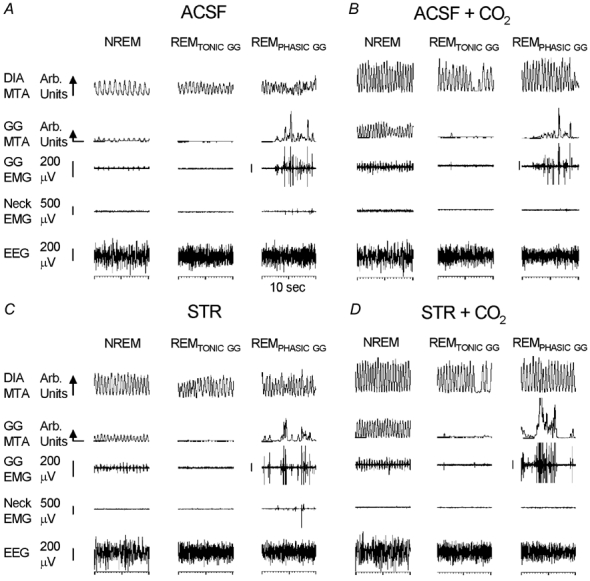
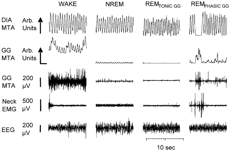
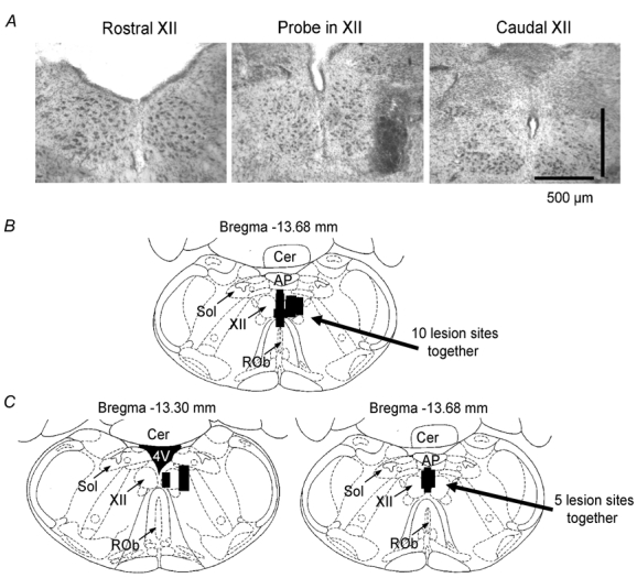
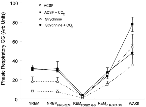
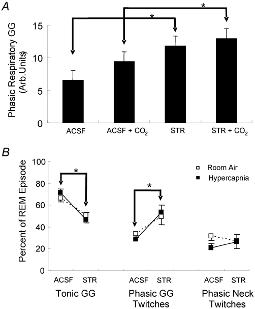
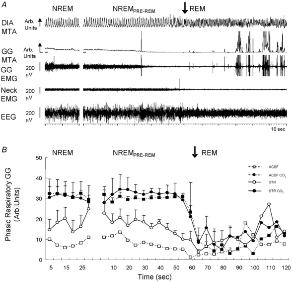
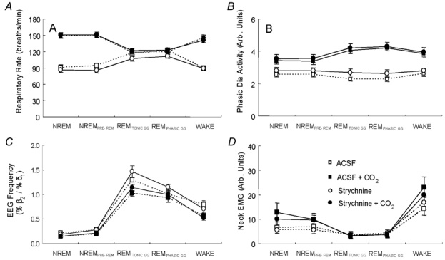
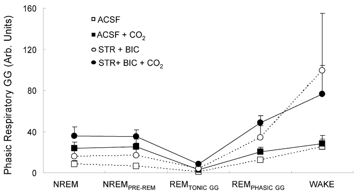
The hypoglossal motor nucleus innervates the genioglossus (GG) muscle of the tongue, a muscle that helps maintain an open airway for effective breathing. Rapid-eye-movement (REM) sleep, however, recruits powerful neural mechanisms that can abolish GG activity even during strong reflex stimulation such as by hypercapnia, effects that can predispose to sleep-related breathing problems in humans. We have developed an animal model to chronically manipulate neurotransmission at the hypoglossal motor nucleus using in vivo microdialysis in freely behaving rats. This study tests the hypothesis that glycine receptor antagonism at the hypoglossal motor nucleus, either alone or in combination with GABAA receptor antagonism, will prevent suppression of GG activity in natural REM sleep during room air and CO2-stimulated breathing. Rats were implanted with electroencephalogram and neck muscle electrodes to record sleep-wake states, and GG and diaphragm electrodes for respiratory muscle recording. Microdialysis probes were implanted into the hypoglossal motor nucleus for perfusion of artificial cerebrospinal fluid (ACSF) and strychnine (glycine receptor antagonist, 0.1 mM) either alone or combined with bicuculline (GABAA antagonist, 0.1 mM) during room air and CO2-stimulated breathing. Compared to ACSF controls, glycine receptor antagonism at the hypoglossal motor nucleus increased respiratory-related GG activity in room air (P = 0.010) but not hypercapnia (P = 0.221). This stimulating effect of strychnine in room air did not depend on the prevailing sleep-wake state (P = 0.625) indicating removal of a non-specific background inhibitory glycinergic tone. Nevertheless, GG activity remained minimal in those REM sleep periods without phasic twitches in GG muscle, with GG suppression from non-REM (NREM) sleep being > 85 % whether ACSF or strychnine was at the hypoglossal motor nucleus or the inspired gas was room air or 7 % CO2. While GG activity was minimal in these REM sleep periods, there was a small but measurable increase in GG activity after strychnine (P < 0.05). GG activity was also minimal, and effectively abolished, in the REM sleep periods without GG twitches with combined glycine and GABAA receptor antagonism at the hypoglossal motor nucleus. We conclude that these data in freely behaving rats confirm that inhibitory glycine and GABAA receptor mechanisms are present at the hypoglossal motor nucleus and are tonically active, but that such inhibitory mechanisms make only a small contribution to the marked suppression of GG activity and reflex responses observed in periods of natural REM sleep.


Figures

Figure 3
Figure 3. GG responses to strychnine at the hypoglossal motor nucleus in natural sleep
Sample traces from one rat showing typical sleep patterns and respiratory muscle activities with microdialysis perfusion of artificial cerebrospinal fluid (ACSF) and strychnine into the hypoglossal motor nucleus during room air and CO2-stimulated breathing. In NREM sleep, strychnine produced increases in respiratory-related genioglossus (GG) activity compared to ACSF in room air (C vs. A) but less so with CO2 (D vs. B). However, GG activity was minimal in periods of REMTONIC GG across all conditions. Abbreviations are as for Fig. 2.
Figure 2
Figure 2. Typical GG activities recorded across sleep–wake states
Shown are the electroencephalogram (EEG), neck electromyogram (EMG), diaphragm (Dia) and genioglossus (GG) signals. The GG and Dia are displayed as their moving-time averages (MTA) in arbitrary (arb) units. The baseline of the integrator (i.e. electrical zero) is shown for the GG MTA. The arrow on the DIA and GG calibration bars denotes the direction of inspiration. Note that despite prominent tonic GG activity during room air breathing in wakefulness, a degree of phasic respiratory-related GG activity is also present. Note also that respiratory-related GG activity is markedly decreased from wakefulness to non-rapid-eye-movement (NREM) sleep, and effectively abolished in those periods of rapid-eye-movement (REM) sleep without phasic GG twitches, i.e. REMTONIC GG. Periods of REM sleep are also associated with phasic bursts of GG activity (i.e. REMPHASIC GG) that did not have a consistent relationship to breathing. Traces are taken during room air breathing and microdialysis perfusion of artificial cerebrospinal fluid into the hypoglossal motor nucleus.
Figure 1
Figure 1. Location of microdialysis probes
A, example of a lesion site made by the microdialysis probe in the hypoglossal motor nucleus. The dark staining is produced by perfusing a microdialysis probe with the membrane cut at the tip with 1 % potassium permanganate to mark the lesion site. Intact cells in the hypoglossal motor nucleus can be seen on this section as well as those rostral (left) and caudal (right). B and C, the distribution of individual microdialysis sites from all rats for studies 1 and 2 respectively. The size of each bar represents the apparent size of the lesion from the histological sections. Abbreviations: AP, area postrema; Cer, cerebellum; 4V, fourth ventricle; Sol, nucleus tractus solitarius; XII, hypoglossal motor nucleus; ROb, raphe obscurus.
Figure 4
Figure 4. Group data showing GG responses to strychnine at the hypoglossal motor nucleus
Group data showing changes in respiratory-related GG activity across all sleep–wake states during microdialysis perfusion of ACSF and strychnine into the hypoglossal motor nucleus during both room air and CO2-stimulated breathing. Note that although GG activity in REMTONIC GG was increased slightly by strychnine, the levels of GG activity in these periods of REM sleep remained minimal during both room air and CO2-stimulated breathing. All values are means ±s.e.m.
Figure 5
Figure 5. Effects of strychnine on GG activity measured over the entire REM episode and the percentage of REM sleep with periods of tonic activity and phasic GG twitches
A, group data showing GG activity measured throughout the entire REM episodes. GG activity was increased with strychnine (STR) at the hypoglossal motor nucleus in REM sleep, compared with ACSF, during both room air and CO2-stimulated breathing. B, group data showing the changes in the proportion of the REM sleep time spent with and without phasic twitches in GG muscle with strychnine and ACSF at the hypoglossal motor nucleus during room air (open symbols) and CO2-stimulated breathing (filled symbols). Responses are also shown for neck muscle activity. The percentage of REM sleep accompanied by phasic GG twitches was increased by strychnine compared with ACSF. All values are means ±s.e.m.*P < 0.05, other abbreviations are as for Fig. 2.
Figure 6
Figure 6. GG activity upon transition from NREM to REM sleep with strychnine at the hypoglossal motor nucleus
A, sample trace from one rat showing the abolition of GG activity at the onset of REM sleep despite strychnine at the hypoglossal motor nucleus. Note the onset of phasic twitches in GG muscle occurred later into the REM sleep episode. Abbreviations are as for Fig. 2. B, group data showing changes in respiratory-related GG activity from NREM to REM sleep during microdialysis perfusion of ACSF and strychnine into the hypoglossal motor nucleus during both room air and CO2-stimulated breathing. GG activity in REM sleep was similar across all conditions. All values are means ±s.e.m. See text for further details.
Figure 7
Figure 7. Group data showing specificity of responses to strychnine at the hypoglossal motor nucleus
Group data showing specificity of responses to strychnine at the hypoglossal motor nucleus. Responses of respiratory rate (A), phasic diaphragm (DIA) activity (B), the ratio of high to low frequencies in the EEG (C) and neck EMG (D) are shown across all sleep–wake states for microdialysis perfusion of ACSF and strychnine into the hypoglossal motor nucleus during both room air and CO2-stimulated breathing. There were no effects of strychnine on any parameter either in room air or hypercapnia. All values are means ±s.e.m.
Figure 8
Figure 8. Effects of combined strychnine and bicuculline on GG activity across sleep–wake states
Group data showing changes in respiratory-related GG activity across all sleep–wake states during microdialysis perfusion of ACSF and combined strychnine and bicuculline into the hypoglossal motor nucleus during both room air and CO2-stimulated breathing. Note that the levels of GG activity were minimal in the periods of REM sleep without phasic GG twitches during both room air and CO2-stimulated breathing. All values are means ±s.e.m.
Figure 9
Figure 9. GG activation by serotonin at the hypoglossal motor nucleus
Example showing an increase in GG activity with microdialysis perfusion of serotonin (5-HT) into the hypoglossal motor nucleus. These traces from one rat were both obtained in NREM sleep. Abbreviations are as for Fig. 2.
Figure 10
Figure 10. Strychnine at the hypoglossal motor nucleus prevents GG responses to glycine at both the ipsilateral and contralateral sides
Microdialysis perfusion of strychnine (STR) into the right hypoglossal motor nucleus (XII) prevented the suppression of both right and left GG activity in response to glycine (GLY) applied to the left motor nucleus. Abbreviations are as for Fig. 2.

Similar articles

Cited by

References

    1. Aston-Jones G, Bloom FE. Activity of norepinephrine-containing locus coeruleus neurons in behaving rats anticipates fluctuations in the sleep-waking cycle. J Neurosci. 1981;1:876–886. - PMC - PubMed
    1. Bailey EF, Jones CL, Reeder JC, Fuller DD, Fregosi RF. Effect of pulmonary stretch receptor feedback and CO2 on upper airway and respiratory pump muscle activity in the rat. J Physiol. 2001;532:525–534. - PMC - PubMed
    1. Bellingham MC, Berger AJ. Presynaptic depression of excitatory synaptic inputs to rat hypoglossal motoneurons by muscarinic M2 receptors. J Neurophysiol. 1996;76:3758–3770. - PubMed
    1. Berger AJ. Determinants of respiratory motoneuron output. Respir Physiol. 2000;122:259–269. - PubMed
    1. Chamberlin NL, Bocchiaro CM, Greene RW, Feldman JL. Nicotinic excitation of rat hypoglossal motoneurons. Neuroscience. 2002;115:861–870. - PubMed

Publication types

MeSH terms

LinkOut - more resources