. 2003 Nov 1;552(Pt 3):933-43.
doi: 10.1113/jphysiol.2003.050062. Epub 2003 Aug 22.

The effects of asphyxia on renal function in fetal sheep at midgestation

Affiliations

The effects of asphyxia on renal function in fetal sheep at midgestation

A E O'Connell et al. J Physiol. .

Abstract

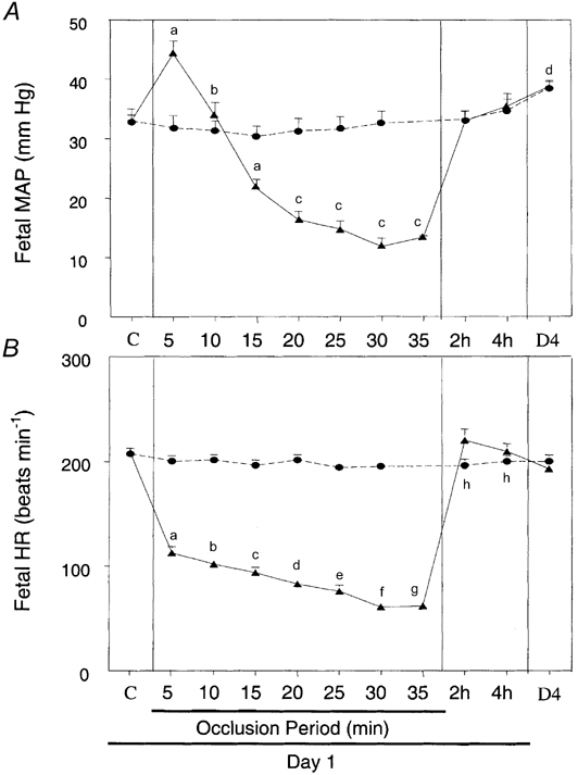
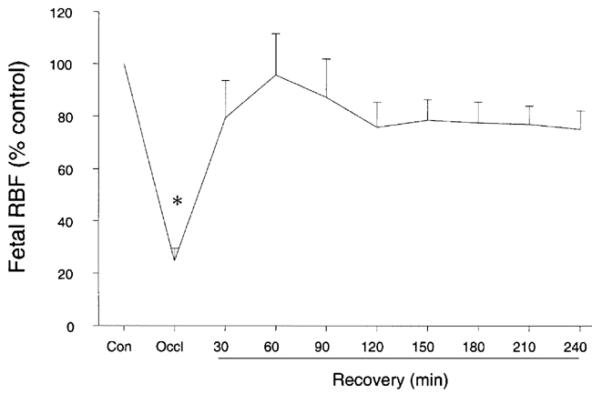
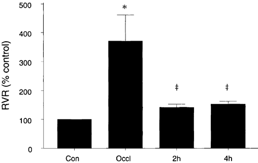
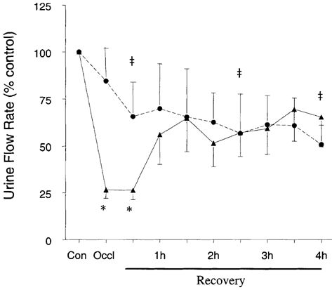
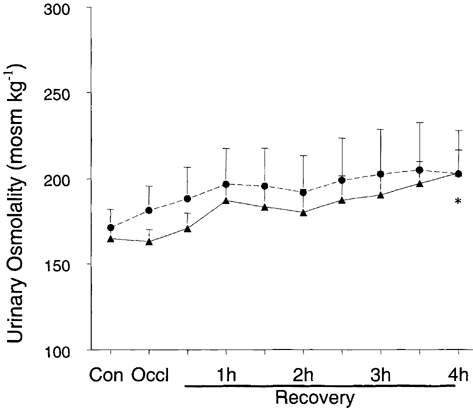
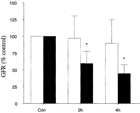
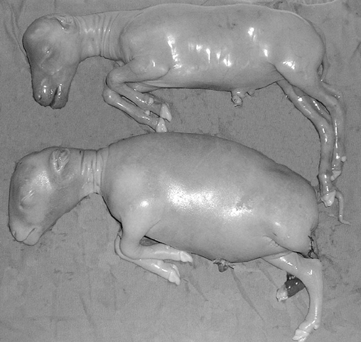
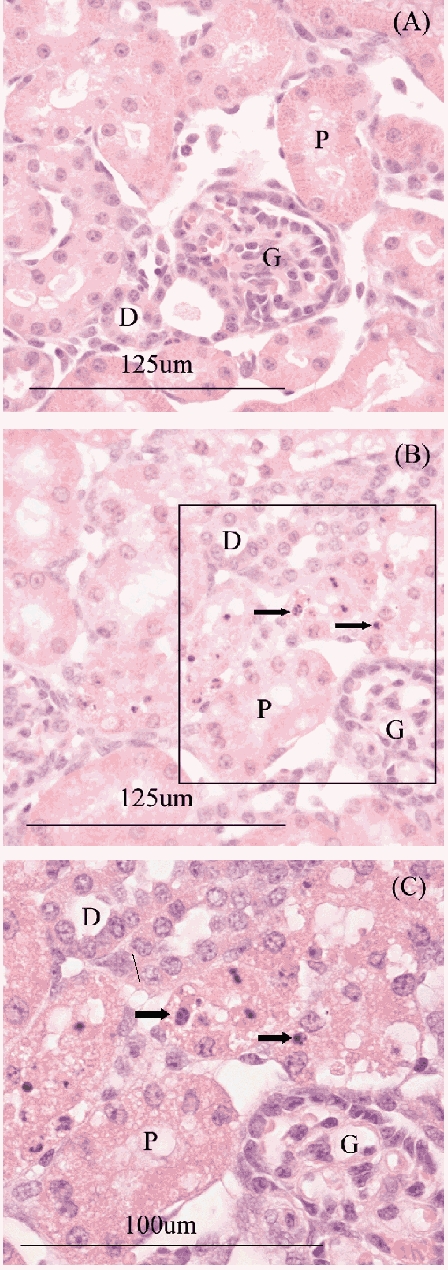
To determine whether damage to the fetal kidneys plays a role in the formation of hydrops fetalis following a severe asphyxial episode, six chronically catheterised fetal sheep, at 0.6 gestation (90 days; term 150 days), were subjected to 30 min of complete umbilical cord occlusion. During the occlusion period, mean arterial pressure, heart rate and renal blood flow decreased (P < 0.001). There were falls in arterial pH and PO2 and a rise in PCO2 (P < 0.001). Urine flow rate decreased (P < 0.005), as did the excretion rates of sodium and osmoles (P < 0.05). However, by 60 min after release of occlusion, urine flow rate was similar to control values. By the end of day 1, most renal variables returned to normal. At post-mortem, 72 h after occlusion, all asphyxiated fetuses showed gross signs of hydrops. Body weight was higher (P < 0.05) due to fluid accumulation in the peritoneal (P < 0.001) and pleural cavities (P < 0.05) as well as subcutaneously (P < 0.05). Amniotic/allantoic fluid volume was increased (P < 0.05). Kidney histology was normal except for clusters of apoptotic cells in some proximal tubules. In conclusion, this severe asphyxial episode caused surprisingly little damage to the kidney and the changes in renal function were very transient. Thus renal damage was not important in the development of hydrops. Possibly, the midgestation fetal kidney has a limited capacity to increase urinary salt and water excretion in response to increased fluid delivery across the placenta.


Figures

Figure 1
Figure 1. Fetal blood pressure and heart rate during and after occlusion
A, fetal mean arterial pressure (MAP) for day 1, including the 30 min occlusion period (35 min for two animals), and day 4. a Different from control and all other occlusion periods, P < 0.05; b different from all occlusion periods, P < 0.001; c different from control, 5, 10 and 15 min, P < 0.05; d different from control, P < 0.05. There was no change in the sham group. B, fetal heart rate (FHR) for day 1, including the 30 min occlusion period (35 min for two animals), and day 4. a Different from control and all other occlusion periods except 10 min, P < 0.001; b different from control and all other occlusion periods except 5 and 15 min, P < 0.025; c different from control and all other occlusion periods except 10 and 20 min P < 0.025; d different from control and all other occlusion periods except 15 and 25 min, P < 0.025; e different from control and all other occlusion periods except 20 min, P < 0.01; f different from control and all other occlusion periods except 35 min, P < 0.05; g different from control and all other occlusion periods except 30 min, P < 0.05; h different from control in sham fetuses, P < 0.05. Sham (•, n = 5), occlusion (▴, n = 6). Values expressed as mean +s.e.m. Occlusion time periods are 5 min averages. Day 4 (D4) compared to control period only.
Figure 2
Figure 2. Fetal renal blood flow (RBF) normalised to control for fetuses subjected to 30 min of umbilical cord occlusion
* Different to all periods, P < 0.001. Values expressed as mean +s.e.m. (n = 5).
Figure 3
Figure 3. Renal vascular resistance relative to control on day 1 in fetuses subjected to 30 min umbilical cord occlusion (n = 5)
* Different from all periods, P < 0.001; ‡ different from control period, P < 0.05. Values expressed as mean +s.e.m.
Figure 4
Figure 4. Plasma concentrations of glucose (A) and lactate (B)
** Different from all periods, P < 0.001, different from control, *P < 0.05; ‡ different from control (Con) and occlusion (Occl), P < 0.05. Sham (open bars) n = 5; occlusion (filled bars) n = 6 except at 4 h, when n = 5. Values expressed as mean +s.e.m.
Figure 5
Figure 5. Urinary flow rate as a percentage of control for midgestational fetal sheep undergoing complete umbilical occlusion (▴, n = 6) or for the sham group (•, n = 5)
* Different from control P < 0.005; ‡ different from control P < 0.05. Values expressed as mean ±s.e.m.
Figure 6
Figure 6. The urinary osmolality, for sham (•; n = 5) and occlusion (▴; n = 6) groups
* Different from control and occlusion periods, P < 0.05. Values expressed as mean +s.e.m.
Figure 7
Figure 7. Glomerular filtration rate (GFR) as a percentage of control for fetuses before and after occlusion
Sham (open bars) n = 5, occlusion (filled bars) n = 6 except at 4 h when n = 5. * Different from control, P < 0.05. Values expressed as mean +s.e.m.
Figure 8
Figure 8
Photograph of an occluded fetus (1270 g, bottom) and its unoperated twin (930 g, top) aged 99 days
Figure 9
Figure 9. Histology slides of the left kidney of a sham fetus (A) and occluded fetus (B)
Note the apoptotic cells in B, arrows, which are shown at higher magnification in C. G, glomerulus; P, proximal tubule; D, distal tubule; arrow, apoptotic cell.

Similar articles

Cited by

References

    1. Alward CT, Hook JB, Helmbrath TA, Bailie MD. Effects of asphyxia on renal function in the newborn piglet. Pediatr Res. 1978;12:225–228. - PubMed
    1. Anderson DF, Faber JJ. Animal model for polyhydramnios. Am J Obstet Gynecol. 1989;160:389–390. - PubMed
    1. Ball RH, Parer JT, Caldwell LE, Johnson J. Regional blood flow and metabolism in ovine fetuses during severe cord occlusion. Am J Obstet Gynecol. 1994;171:1549–1555. - PubMed
    1. Bennet L, Rossenrode S, Gunning MI, Gluckman PD, Gunn AJ. The cardiovascular and cerebrovascular responses of the immature fetal sheep to acute umbilical cord occlusion. J Physiol. 1999;517:247–257. - PMC - PubMed
    1. Blair DK, Vander Straten MC, Gest AL. Hydrops in fetal sheep from rapid induction of anemia. Pediatr Res. 1994;35:560–564. - PubMed

Publication types

MeSH terms

LinkOut - more resources