

In vivo determination of steric and electrostatic exclusion of albumin in rat skin and skeletal muscle
- PMID: 12937287
- PMCID: PMC2343468
- DOI: 10.1113/jphysiol.2003.049379
In vivo determination of steric and electrostatic exclusion of albumin in rat skin and skeletal muscle
Abstract
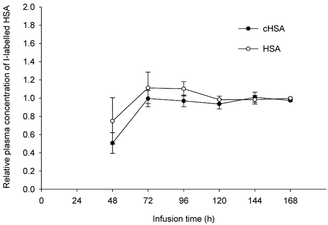
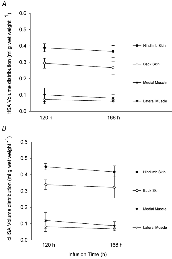
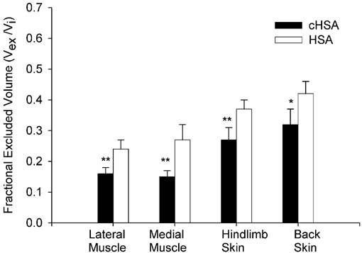
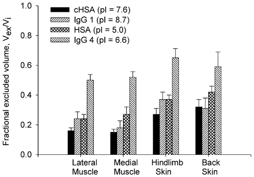
In order to estimate the magnitude of electrostatic exclusion provided by the fixed negative charges of the skin and muscle interstitia of rat in vivo we measured the distribution volumes of two differently charged albumin probes within these tissues. An implanted osmotic pump was used to reach and maintain a steady-state extracellular concentration of a mixture containing two iodine-labelled probes: a charged-modified human serum albumin, cHSA (i.e. a positive probe, isoelectirc point (pI) = 7.6) and a native human serum albumin, HSA (i.e. a normally charged, negative probe, pI = 5.0). Steady-state tissue concentrations were achieved after intravenous infusion of probes for 5-7 days. At the end of this period the animals were nephrectomized and a bolus of 51Cr-EDTA was administered for estimating the extracellular volume. Plasma volumes were measured as 5-min distribution volume of 125I-HSA in separate experiments. The steady-state interstitial fluid concentrations of all probes were determined using nylon wicks implanted postmortem. Calculations of labelled probes were made for interstitial fluid volumes (Vi), extravascular albumin distribution volumes (Vav,a) and relative interstitial excluded volume fractions (Vex,a/Vi). We found that the positive probe is excluded from a significantly smaller fraction of the interstitium. Specifically, the average relative albumin exclusion fractions obtained were: 16% and 26% in skeletal muscle and 30% and 40% in skin, for cHSA and HSA, respectively. On average, the fixed negative charges of the interstitium are responsible for about 40% of the total albumin exclusion in skeletal muscle and 25% in the whole skin tissue and thus, contribute significantly to volume exclusion in these tissues.
Figures
Comment in
-
Intersitial exluded volumes: the effect of charge.J Physiol. 2003 Dec 1;553(Pt 2):333. doi: 10.1113/jphysiol.2003.053595. Epub 2003 Oct 24. J Physiol. 2003. PMID: 14578482 Free PMC article. No abstract available.
Similar articles
-
The interstitial distribution of macromolecules in rat tumours is influenced by the negatively charged matrix components.J Physiol. 2005 Sep 1;567(Pt 2):557-67. doi: 10.1113/jphysiol.2005.089615. Epub 2005 Jun 30. J Physiol. 2005. PMID: 15994186 Free PMC article.
-
Effect of charge on interstitial distribution of albumin in rat dermis in vitro.J Physiol. 2003 Jul 15;550(Pt 2):505-14. doi: 10.1113/jphysiol.2003.042713. Epub 2003 May 23. J Physiol. 2003. PMID: 12766239 Free PMC article.
-
Interstitial exclusion of albumin in rat tissues measured by a continuous infusion method.Am J Physiol. 1992 Oct;263(4 Pt 2):H1222-33. doi: 10.1152/ajpheart.1992.263.4.H1222. Am J Physiol. 1992. PMID: 1415771
-
Effect of hydration on interstitial distribution of charged albumin in rat dermis in vitro.J Physiol. 2005 Dec 1;569(Pt 2):631-41. doi: 10.1113/jphysiol.2005.096792. Epub 2005 Oct 6. J Physiol. 2005. PMID: 16210353 Free PMC article.
-
Interstitial exclusion of positively and negatively charged IgG in rat skin and muscle.Am J Physiol Heart Circ Physiol. 2001 Apr;280(4):H1505-12. doi: 10.1152/ajpheart.2001.280.4.H1505. Am J Physiol Heart Circ Physiol. 2001. PMID: 11247760
Cited by
-
Intersitial exluded volumes: the effect of charge.J Physiol. 2003 Dec 1;553(Pt 2):333. doi: 10.1113/jphysiol.2003.053595. Epub 2003 Oct 24. J Physiol. 2003. PMID: 14578482 Free PMC article. No abstract available.
-
The interstitial distribution of macromolecules in rat tumours is influenced by the negatively charged matrix components.J Physiol. 2005 Sep 1;567(Pt 2):557-67. doi: 10.1113/jphysiol.2005.089615. Epub 2005 Jun 30. J Physiol. 2005. PMID: 15994186 Free PMC article.
-
Albumin is an interface between blood plasma and cell membrane, and not just a sponge.Clin Kidney J. 2021 Oct 5;15(4):624-634. doi: 10.1093/ckj/sfab194. eCollection 2022 Apr. Clin Kidney J. 2021. PMID: 35371452 Free PMC article.
-
Body fluid dynamics: back to the future.J Am Soc Nephrol. 2011 Dec;22(12):2166-81. doi: 10.1681/ASN.2011080865. Epub 2011 Oct 27. J Am Soc Nephrol. 2011. PMID: 22034644 Free PMC article. Review.
-
Cytokine signalling in rat pulp interstitial fluid and transcapillary fluid exchange during lipopolysaccharide-induced acute inflammation.J Physiol. 2006 May 15;573(Pt 1):225-36. doi: 10.1113/jphysiol.2006.104711. Epub 2006 Mar 9. J Physiol. 2006. PMID: 16527857 Free PMC article.
References
-
- Aukland K, Fadnes HO. Protein concentration of interstitial fluid collected from rat skin by wick method. Acta Physiol Scand. 1973;88:350–358. - PubMed
-
- Aukland K, Reed RK. Interstitial-lymphatic mechanisms in the control of extracellular volume. Physiol Rev. 1993;73:1–78. - PubMed
-
- Bell DR. Extravascular transport and distribution of charge-modified albumin in skin (Abstract) Microvasc Res. 1985;29:207.
-
- Bert JL, Pearce RH. Handbook of Physiology. The Cardiovascular System. Microcirculation. IV. Bethesda, MD: Blackwell Science Inc; 1984. The interstitium and microvascular exchange; pp. 521–547. Section 2, Part 1, Chapter 12, pp.
Publication types
MeSH terms
Substances
LinkOut - more resources
Full Text Sources
Other Literature Sources
Research Materials
Miscellaneous