. 2003 Nov 1;552(Pt 3):727-41.
doi: 10.1113/jphysiol.2003.049312. Epub 2003 Aug 22.

Contribution of Ca2+-dependent conductances to membrane potential fluctuations of medullary respiratory neurons of newborn rats in vitro

Affiliations

Contribution of Ca2+-dependent conductances to membrane potential fluctuations of medullary respiratory neurons of newborn rats in vitro

Hiroshi Onimaru et al. J Physiol. .

Abstract

Ca2+-dependent conductances were studied in respiratory interneurons in the brainstem-spinal cord preparation of newborn rats. omega-Conotoxin-GVIA attenuated evoked postsynaptic potentials, spontaneous or evoked inspiratory spinal nerve activity and blocked spike afterhyperpolarization. Furthermore, omega-conotoxin-GVIA augmented rhythmic drive potentials of pre-inspiratory and inspiratory neurons and increased respiratory-related spike frequency of pre-inspiratory cells with no effect on inspiratory hyperpolarization. In contrast, omega-agatoxin-IVA depressed drive potentials of pre-inspiratory and inspiratory neurons and attenuated inspiratory hyperpolarization and spike frequency of pre-inspiratory cells. It did not affect spike shape and exerted only minor, non-significant, attenuating effects on spontaneous or evoked nerve bursts or evoked postsynaptic potentials. Nifedipine diminished drive potentials and spike frequency of pre-inspiratory neurons and shortened drive potentials in some cells. omega-Conotoxin-MVIIC attenuated drive potentials and intraburst firing rate of pre-inspiratory neurons and decreased substantially respiratory frequency. Respiratory rhythm disappeared following combined application of omega-conotoxin-GVIA, omega-conotoxin-MVIIC, omega-agatoxin-IVA and nifedipine. Apamin potentiated drive potentials and abolished spike afterhyperpolarization, whereas charybdotoxin and tetraethylammonium prolonged spike duration without effect on shape of drive potentials. The results show that specific sets of voltage-activated L-, N- and P/Q-type Ca2+ channels determine the activity of particular subclasses of neonatal respiratory neurons, whereas SK- and BK-type K+ channels attenuate drive potentials and shorten spikes, respectively, independent of cell type. We hypothesize that modulation of spontaneous activity of pre-inspiratory neurons via N-, L- and P/Q-type Ca2+ channels is important for respiratory rhythm or pattern generation.


Figures
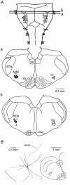
Figure 1
Figure 1. Location of recorded neurons
A, distribution of recorded neonatal ventral respiratory group (VRG) neurons of the ventrolateral medulla (VLM). Pre-inspiratory (Pre-I, filled circles) and inspiratory (Insp, open circles) VLM-VRG neurons were recorded within ±100 μm distance from the lines (a and b) indicating the rostrocaudal position of cross-sections. Cells are plotted to distinct sides of the medulla for higher clarity. XII, hypoglossal nucleus; STT, spinal trigeminal tract; STN, spinal trigeminal nucleus; AMB, ambigual nucleus; IO, inferior olive; RFN, retrofacial nucleus; CST, corticospinal tract. B, histological analysis of a Lucifer-Yellow-filled cell showed that the soma, most portions of the dendritic tree and the axon were located at a depth > 200 μm from the ventral surface. In this cell it was found that apamin (0.4 μm) abolished the afterhyperpolarization of single spikes within 12 min after the start of superfusion of the drug. A very similar distribution of cell processes and kinetics of drug effects was observed for > 70 % of recorded neurons. (For details, see text.)
Figure 2
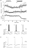
Figure 2. Effects of ω-conotoxin-GVIA (ω-Cono-GVIA) on a Pre-I neuron
A, time course of changes in the respiratory drive potential amplitude (Vm Burst, circles), burst interval (Interval, squares) of Pre-I neuron activity and amplitude of spinal nerve burst (C4, triangles) induced by ω-Cono-GVIA (2 μm). B, traces of membrane potential (Vm) of the Pre-I neuron and of C4 activity; a and b correspond to a and b in A, respectively. Note that the toxin did not affect inspiratory-related inhibition, whereas the amplitude of the drive potential increased to about 150 % of control. C, potentiating effect of ω-Cono-GVIA on respiratory drive potential amplitude. The recording shows at higher time resolution a single respiratory-related burst indicating how the relative changes in drive potential amplitude and spike frequencies of Table 2 were determined. Left, control close to a in A. Right, during ω-Cono-GVIA close to b in A.
Figure 5
Figure 5. Effects of Ca2+ channel blockers on an Insp-III neuron
A, time course of changes in drive potential amplitude (Vm Burst, circles) as well as burst interval (Interval, squares) of Insp-III neuron activity, and amplitude of spinal nerve burst (C4, triangles) upon consecutive administration of nifedipine (Nif, 20 μm), ω-Cono-GVIA (2 μm) and ω-Aga-IVA (0.2 μm). Augmentation of inspiration-related neuronal drive potential induced by ω-Cono-GVIA was reversed into a decline of the response by ω-Aga-IVA. Note that the C4 amplitude was reduced to about 50 % of control by ω-Cono-GVIA. B, individual oscillations of membrane potential (Vm) of the Insp-III neuron and of C4 activity; a-f correspond to time periods a-f in A, respectively.
Figure 4
Figure 4. Effects of ω-Aga-IVA on a Pre-I neuron
A, time course of changes in drive potential amplitude (Vm Burst, circles) as well as burst interval (Interval, squares) of Pre-I neuron activity and cervical nerve burst amplitude (C4, triangles) due to ω-Aga-IVA (0.2 μm). Note that burst amplitude was reduced to < 50 % of control. B, traces of membrane potential (Vm) of Pre-I neuron and C4 activity; a and b correspond to time periods a and b in A, respectively. Note in b that attenuation of neuronal activity was accompanied by a failure of nerve discharge.
Figure 3
Figure 3. Effects of ω-agatoxin-IVA (ω-Aga-IVA) and ω-Cono-GVIA on an inspiratory type-III (Insp-III) neuron
A, time course of changes in drive potential amplitude (Vm Burst, circles) and burst interval (Interval, squares) of neuronal activity and spinal nerve burst amplitude (C4, triangles) upon successive administration of ω-Aga-IVA (0.2 μm) and ω-Cono-GVIA (2 μm). Note that ω-Aga-IVA irreversibly reduced the drive potential amplitude to about 60 % of control, whereas the amplitude of single C4 nerve bursts only decreased slightly. The inspiration-related drive potential of the neuron re-increased upon subsequent addition of ω-Cono-GVIA, but C4 activity declined further to < 50 % of control. B, individual neuronal and nerve bursts at time periods a-c in A. C, action potentials induced by a 10 ms depolarizing current pulse at time periods a-c in A. Note the decrease in spike afterhyperpolarization after ω-Cono-GVIA.
Figure 6
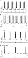
Figure 6. Effects of ω-Cono-MVIIC on respiratory neurons
A and B, effects on a Pre-I neuron. A, control. B, 16 min after start of exposure to 2 μmω-Cono-MVIIC. Bath-application of the agent resulted in a substantial decrease of respiratory frequency that was not accompanied by a decrease in burst frequency of the Pre-I cell. Note that ω-Cono-MVIIC reduced drive potential amplitude. C and D, effects on an Insp-III neuron. C, control. D, 15 min after start of exposure to 2 μmω-Cono-MVIIC. The drug depressed the frequency of rhythmic activity in the same way as it slowed inspiratory nerve activity. Note that the pre- and post-inspiratory hyperpolarizations due to activity of presynaptic Pre-I cells were uncoupled from inspiratory activity.
Figure 7
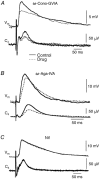
Figure 7. Effects of Ca2+ channel blockers on postsynaptic potentials (PSPs) and nerve activity evoked by electrical stimulation of the contralateral ventrolateral medulla
Vm, membrane potential, C4, spinal nerve reflex. Continuous line, control responses before drug application; dashed line, responses 20–30 min after start of drug application. A, effect of ω-Cono-GVIA (2 μm) on an Insp-III neuron. B, effect of ω-Aga-IVA (0.2 μm) on an Insp-I neuron. C, effect of Nif (20 μm) on an Insp-I neuron. Each trace represents average of 10 (A) or 6 (B and C) sweeps.
Figure 8
Figure 8. Effects of cocktail application of Ca2+ channel blockers on bursting in an Insp-II neuron
A, time course of changes in the drive potential amplitude (ΔVm, open circles), burst duration (Duration, downward triangles) as well as burst interval (Interval, squares) of Insp-II neuron activity and amplitude of spinal nerve burst (C4, upward triangles) upon simultaneous administration of Nif (20 μm), ω-Cono-GVIA (3 μm), ω-Cono-MVIIC (3 μm) and ω-Aga (0.2 μm). Amplitude of evoked EPSPs (ΔVm) by stimulation of the contralateral vetrolateral medulla is also plotted (filled circles). Inspiration-related neuronal drive potential amplitude, burst duration and C4 nerve burst amplitude irreversibly decreased, whereas the burst interval increased even after wash out. B, traces of membrane potential (Vm) of the Insp-II neuron and C4 activity that correspond to time periods a-c in A, respectively. C, evoked EPSPs (average of 5 traces) of the Insp-II neuron and C4 activity that correspond to time periods a-c in A, respectively.
Figure 9
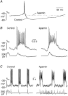
Figure 9. Effects of apamin on burst activity and spike afterhyperpolarization
A, in a Pre-I neuron, apamin (4 μm) irreversibly suppressed afterhyperpolarization of a spontaneous single action potential after 5 min. Each trace represents the mean of 4 sweeps. B, in an Insp-III cell, apamin (0.4 μm) potentiated inspiratory-related drive potential after 8 min, whereas pre- and post-inspiratory hyperpolarizations were not profoundly affected. C, in a Pre-I neuron, potentiation of respiratory-related drive potential seen 8 min after application of apamin (1 μm) was associated with inactivation of spike discharge. In contrast, apamin had no major effect on inspiratory-related hyperpolarization and spike inhibition. Note that regular bursting of both neurons was not perturbed by the bee venom despite occurrence of massive irregular nerve (C4) bursting.
Figure 11
Figure 11. Effects of charybdotoxin on spikes and burst activity
A, in an Insp-II cell, charybdotoxin (0.2 μm) increased duration of spontaneous spikes by ≈50 % after 30 min of exposure. Each trace represents the mean of 6 sweeps. B and C, in a Pre-I neuron, combined administration of charybdotoxin (0.2 μm, 30 min preincubation) and apamin (0.5 μm, 15 min preincubation) did not perturb rhythmic bursting despite massive disturbance of spinal nerve (C4) discharge. Note that this cell did not respond with an increased burst potential to apamin (for details, see text).
Figure 10
Figure 10. Effects of tetraethylammonium (TEA) on individual spikes and burst activity
A, in a Pre-I neuron TEA (1 mm) increased duration of single spontaneous action potentials by > 50 %. Each trace represents average of 7 sweeps. B and C, TEA (1 mm) did not change membrane potential (Vm) fluctuations of a Pre-I neuron within 15 min of administration despite occurrence of tonic activity in spinal nerve recording (C4).

Similar articles

Cited by

References

    1. Arata A, Onimaru H, Homma I. Respiration-related neurons in the ventral medulla of newborn rats in vitro. Brain Res Bull. 1990;24:599–604. - PubMed
    1. Ballanyi K, Onimaru H, Homma I. Respiratory network function in the isolated brainstem-spinal cord of newborn rats. Prog Neurobiol. 1999;59:583–634. - PubMed
    1. Bianchi AL, Denavit-Saubie M, Champagnat J. Central control of breathing in mammals: neuronal circuitry, membrane properties, and neurotransmitters. Physiol Rev. 1995;75:1–45. - PubMed
    1. Brockhaus J, Ballanyi K. Synaptic inhibition in the isolated respiratory network of neonatal rats. Eur J Neurosci. 1998;10:3823–3839. - PubMed
    1. Brockhaus J, Ballanyi K. Anticonvulsant A1 receptor-mediated adenosine action on neuronal networks in the brainstem-spinal cord of newborn rats. Neuroscience. 2000;96:359–371. - PubMed

Publication types

LinkOut - more resources