. 2003 Nov 1;552(Pt 3):833-44.
doi: 10.1113/jphysiol.2003.048165. Epub 2003 Aug 22.

Insulin-like growth factor-1 prevents age-related decrease in specific force and intracellular Ca2+ in single intact muscle fibres from transgenic mice

Affiliations

Insulin-like growth factor-1 prevents age-related decrease in specific force and intracellular Ca2+ in single intact muscle fibres from transgenic mice

Estela Gonzalez et al. J Physiol. .

Abstract

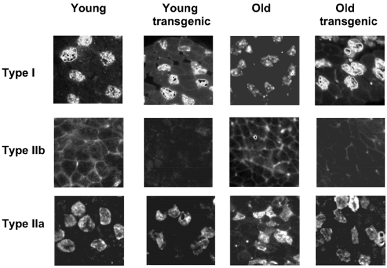
In the present work we test the hypothesis that sustained transgenic overexpression of insulin-like growth factor-1 (IGF-1) in skeletal muscle prevents age-related decreases in myoplasmic Ca2+ concentration and consequently in specific force in single intact fibres from the flexor digitorum brevis (FDB) muscle from the mouse. Measurements of IGF-1 concentration in FDB muscle showed higher levels in transgenic than in wild-type mice at all ages. The specific tetanic force decreased significantly in single muscle fibres from old (286 +/- 22 kPa) compared to young wild-type (455 +/- 28 kPa), young transgenic (423 +/- 43 kPa), and old transgenic mice (386 +/- 15 kPa) (P < 0.05). These results are consistent with measurements in whole FDB muscles. The peak Ca2+ concentration values in response to prolonged stimulation were: 1.47 +/- 0.15, 1.70 +/- 0.29, 0.97 +/- 0.13 and 1.7 +/- 0.22 microM, in fibres from young wild-type, young transgenic, old wild-type and old transgenic mice, respectively. The effects of caffeine on FDB fibres support the conclusion that the age-related decline in peak myoplasmic Ca2+ and specific force is not explained by sarcoplasmic reticulum Ca2+ depletion. Immunohistochemistry in muscle cross-sections was performed to determine whether age and/or IGF-1 overexpression induce changes in fibre type composition. The relative percentages of type IIa, IIx and I myosin heavy chain (MHC) isoforms did not change significantly with age or genotype. Therefore, IGF-1 prevents age-related decline in peak intracellular Ca2+ and specific force in a muscle that does not exhibit changes in fibre type composition with senescence.


Figures

Figure 1
Figure 1. Maximum specific force in FDB muscles from young adult and old, wild-type and IGF-1 transgenic mice
Tetanic contraction declined significantly in old wild-type compared to young, young transgenic and old IGF-1 transgenic mice (P < 0.05). No statistically significant differences were detected among young, young transgenic and old transgenic(P > 0.05). Twitch specific force was not significantly different among groups. #, statistically significant. Data values are represented as the mean +s.e.m.
Figure 2
Figure 2. Force-frequency and [Ca2+]-frequency relationships in single intact FDB muscle fibres from young and old wild-type and IGF-1 transgenic mice
Tension (A) and myoplasmic Ca2+ concentration (B) have been normalized to the maximum value for each fibre. Data points are expressed as means ±s.e.m. Results have been fitted to a single exponential function. No statistically significant difference was detected among groups (P > 0.05).
Figure 4
Figure 4. Maximum specific twitch force, tetanic force, and peak intracellular Ca2+ concentration in single intact FDB muscle fibres from young adult and old wild-type and IGF-1 transgenic mice
A, tetanic contraction declined significantly in old wild-type compared to young wild-type and transgenic and old transgenic mice (P < 0.05). No significant differences in twitch specific force were detected among the four groups. B, maximum intracellular Ca2+ concentration recorded in response to twitch or tetanic pulses in intact FDB fibres. #, statistically significant difference between the old wild-type and the other three groups (P < 0.05). No statistically significant difference was detected among young, young transgenic, and old transgenic mice. Data values are represented as means +s.e.m.
Figure 3
Figure 3. Contraction and intracellular Ca2+ recordings in single intact FDB fibres
Representative records of twitch (A) and tetanic (B) contractions and intracellular Ca2+. Traces in a and b correspond to contraction and intracellular Ca2+ transients, respectively, recorded in single intact FDB fibres from young and old wild-type and transgenic mice. Both maximum specific force and peak intracellular Ca2+ concentration are significantly lower in old wild-type than in young or old transgenic mice. Fibre tension is expressed as normalized values to cross-sectional area, in kilopascals. Ca2+ concentration is expressed in absolute values, as explained in Methods. Contraction and intracellular Ca2+ transients have the same time scale. Dashed lines indicate the force and myoplasmic Ca2+ baselines.
Figure 5
Figure 5. Effects of caffeine on maximum specific force and intracellular Ca2+ concentration in single intact fibres from young and old wild-type and transgenic mice
Specific force (A) and peak myoplasmic Ca2+ concentration (B) before and after 5 mm caffeine (within 30 min incubation). * and #, statistical significance within groups (before and after caffeine) and between old wild-type mice and the remaining groups before caffeine (P < 0.05), respectively. **, statistically significant difference between muscle fibres from young and old wild-type mice treated with caffeine.
Figure 6
Figure 6. Fibre-type composition in ageing and IGF-1 transgenic FDB muscle
Representative serial sections of FDB muscle immunostained with labelled antibodies for type I, IIa and IIb fibres from young and old wild-type and IGF-1 transgenic mice. Quantification of the relative percentages of muscle fibre subtypes is shown in Table 2.

Similar articles

Cited by

References

    1. Allen DG, Duty S, Westerblad H. Metabolic changes in muscle during exercise; their effects on muscle function. Proc Aust Physiol Pharmacol Soc. 1993;24:65–75.
    1. Allen DG, Westerblad H. The effects of caffeine on intracellular calcium, force and the rate of relaxation of mouse skeletal muscle. J Physiol. 1995;487:331–342. - PMC - PubMed
    1. Ashley CC, Mulligan IP, Lea TJ. Ca2+ and activation mechanisms in skeletal muscle. Q Rev Biophys. 1991;24:1–73. - PubMed
    1. Bakker AJ, Head SI, Stephenson DG. Time course of calcium transients derived from Fura-2 fluorescence measurements in single fast twitch fibres of adult mice and rat myotubes developing in primary culture. Cell Calcium. 1997;21:359–364. - PubMed
    1. Barton-Davis ER, Shoturma DI, Musaro A, Rosenthal N, Sweeney HL. Viral mediated expression of insulin-like growth factor I blocks the aging-related loss of skeletal muslce function. Proc Natl Acad Sci U S A. 1998;95:15603–15607. - PMC - PubMed

Publication types

MeSH terms

LinkOut - more resources