. 2003 Nov 1;552(Pt 3):809-21.
doi: 10.1113/jphysiol.2003.047944. Epub 2003 Aug 22.

P2X2 subunits contribute to fast synaptic excitation in myenteric neurons of the mouse small intestine

Affiliations

P2X2 subunits contribute to fast synaptic excitation in myenteric neurons of the mouse small intestine

Jianhua Ren et al. J Physiol. .

Abstract

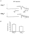
P2X receptors are ATP-gated cation channels composed of one or more of seven different subunits. ATP acts at P2X receptors to contribute to fast excitatory postsynaptic potentials (fEPSPs) in myenteric neurons but the subunit composition of enteric P2X receptors is unknown. These studies used tissues from P2X2 wild-type (P2X2+/+) and P2X2 gene knockout (P2X2-/-) mice to investigate the role of this subunit in enteric neurotransmission. Intracellular electrophysiological methods were used to record synaptic and drug-induced responses from ileal myenteric neurons in vitro. Drug-induced longitudinal muscle contractions and peristaltic contractions of ileal segments were also studied in vitro. Gastrointestinal transit was measured as the progression in 30 min of a liquid radioactive marker administered by gavage to fasted mice. RT-PCR analysis of mRNA from intestinal tissues and data from immunohistochemical studies verified P2X2 gene deletion. The fEPSPs recorded from S neurons in tissues from P2X2+/+ mice were reduced by mecamylamine (nicotinic cholinergic receptor antagonist) and PPADS (P2X receptor antagonist). The fEPSPs recorded from S neurons from P2X2-/- mice were unaffected by PPADS but were blocked by mecamylamine. ATP depolarized S and AH neurons from P2X2+/+ mice. ATP depolarized AH but not S neurons from P2X2-/- mice. alpha,beta-Methylene ATP (alpha,beta-mATP)(an agonist at P2X3 subunit-containing receptors) did not depolarize S neurons but it did depolarize AH neurons in P2X2+/+ and P2X2-/- mice. Peristalsis was inhibited in ileal segments from P2X2-/- mice but longitudinal muscle contractions caused by nicotine and bethanechol were similar in segments from P2X2+/+ and P2X2-/- mice. Gastrointestinal transit was similar in P2X2+/+ and P2X2-/- mice. It is concluded that P2X2 homomeric receptors contribute to fEPSPs in neural pathways underlying peristalsis studied in vitro.


Figures

Figure 1
Figure 1. Representative RT-PCR data showing the presence of P2X2 subunit transcripts in P2X2+/+ tissues but not in P2X2−/− tissues
RT-PCR was performed on whole ileum segments (I), ileal myenteric plexus (IMP), whole colon segments (C) and colonic myenteric plexus (CMP) from 2 separate P2X2+/+ and P2X2−/− mice (data shown from one each). GAPDH, P2X2 and P2X3 autoradiograms were exposed for 15 min, 15 min and 5 h, respectively.
Figure 2
Figure 2. Immunohistochemical localization of the P2X2 subunit in the mouse small intestinal myenteric plexus
A, P2X2 immunoreactivity is present in myenteric neurons in a whole mount preparation taken from the ileum of a P2X2+/+ mouse. Most neurons express P2X2 subunit immunoreactivity. B, P2X2 immunoreactivity is absent from the ileal myenteric plexus of P2X2−/− mice. Scale bar applies to both panels.
Figure 3
Figure 3. P2X2 gene deletion does not alter the action potential or the action potential afterhyperpolarization in myenteric AH neurons
Upper traces are recordings obtained from an AH neuron in the myenteric plexus from a P2X2+/+ mouse. The traces show a long-lasting afterhyperpolarization (left) that follows a single action potential shown on the right on an expanded time scale. Lower traces traces show similar recordings obtained from an AH neuron in the myenteric plexus from a P2X2−/− mouse. There were no differences in the afterhyperpolarization or the action potential recorded from tissues in the two types of mice (see text for details).
Figure 4
Figure 4. P2X2 gene deletion does not alter the action potential in S neurons
Upper traces show that the action potential recorded from S neurons is followed by a brief afterhyperpolarization (arrows) in the myenteric plexus from P2X2+/+ (left) and P2X2−/− (right) mice. Middle traces show that P2X2 subunit gene deletion does not alter the action potential in S neurons. The bottom traces show that the action potential in S neurons is blocked by tetrodotoxin.
Figure 6
Figure 6. Mecamylamine reduced fEPSPs in S neurons from P2X2+/+ mice but completely blocked fEPSPs in S neurons from P2X2−/− mice
A, representative fEPSPs recorded from S neurons in P2X2+/+ (left) and P2X2−/− (right) tissues. The fEPSP in P2X2+/+ neurons is only partly inhibited by mecamylamine (10 μm), but is blocked by mecamylamine in neurons from P2X2−/− mice. B, mean data showing that mecamylamine caused a partial, but significant, reduction in the fEPSP amplitude in P2X2+/+ neurons (n = 5). Mecamylamine completely inhibited the fEPSP in P2X2−/− neurons (n = 9). * Significantly different from P2X2+/+ or P2X2−/− control amplitude (P < 0.05); # Significantly different from the amplitude of the P2X2+/+ fEPSP in the presence of mecamylamine (P < 0.05).
Figure 5
Figure 5. PPADS inhibits fEPSPs recorded from S neurons in tissues from P2X2+/+ but not P2X2−/− mice
A, representative fEPSPs recorded from S neurons in P2X2+/+ (left) and P2X2−/− (right) tissues in the absence or presence of PPADS (10 μm). The fEPSPs recorded from neurons in tissues from P2X2+/+ mice were inhibited by PPADS (left) while fEPSPs recorded from neurons in P2X2−/− tissues were unaffected by PPADS (right). B, mean data showing that the amplitudes of fEPSPs recorded from S neurons in P2X2+/+ (n = 5) and P2X2−/− (n = 6) tissues were similar. PPADS did not alter fEPSP amplitude recorded from P2X2−/− S neurons but it did inhibit the fEPSP in S neurons from P2X2+/+ tissues. * Significantly different from P2X2+/+ control (P < 0.05).
Figure 7
Figure 7. Agonist-induced depolarizations of S neurons in the myenteric plexus from P2X2+/+ or P2X2−/− mice
A, nicotine caused similar amplitude depolarizations of S neurons from P2X2+/+ and P2X2−/− mice. ATP depolarized S neurons from P2X2+/+ but not P2X2−/− mice, while α,β-mATP did not depolarize any S neuron. Drugs were applied (at the arrows) by pressure ejection from a pipette positioned near the impaled neurons. The concentration of each drug in the pipette was 1 mm. B, mean data from experiments similar to those shown in A. There was no difference in the amplitude of the nicotine-induced depolarization recorded from S neurons in tissues from P2X2+/+ (n = 15) and P2X2−/− (n = 12) mice. * The ATP-induced depolarization was significantly smaller in P2X2−/− neurons compared to those in recorded from P2X2+/+ neurons. α,β-mATP did not elicit a response in S neurons from either type of mouse.
Figure 8
Figure 8. AH neurons from P2X2+/+ and P2X2−/− mice were depolarized by α,β-mATP
A, α,β-mATP was applied by pressure ejection (at the arrows) from a pipette positioned near the impaled neuron. The concentration of α,β-mATP in the pipette was 1 mm. B, data showing that the amplitude of the α,β-mATP-induced depolarization was similar in tissues from P2X2+/+ (n = 3) and P2X2−/− (n = 4) mice.
Figure 9
Figure 9. Peristalsis is impaired in ileal segments from P2X2−/− mice
A, pressure-related increases in the peak amplitude of peristaltic contractions. The peak amplitude in tissues from P2X2−/− mice (n = 7) was smaller than that in P2X2+/+ mice (n = 7) (*P < 0.05). Mecamylamine (10 μm) reduced the peak contractions in tissues from P2X2+/+ and P2X2−/− mice (#P < 0.05). B, pressure-related increases in contraction frequency were reduced in tissues from P2X2−/− mice (*P < 0.05). Mecamylamine reduced contraction frequency in tissues from P2X2+/+ and P2X2−/− mice (#P < 0.05).
Figure 10
Figure 10. PPADS inhibits peristalsis in ileal segments from P2X2+/+ but not P2X2−/− mice
A, the peak amplitude of phasic contractions was smaller in tissues from P2X2−/− mice compared to that in P2X2+/+ mice (*P < 0.05) but PPADS (10 μm) did not reduce contraction amplitude in P2X2−/− tissues. PPADS reduced the amplitude of phasic contractions in tissues from P2X2+/+ mice (#P < 0.05). B, the frequency of pressure-induced phasic contractions was lower in P2X2−/− tissues (*P < 0.05) and PPADS did not reduce further the contraction frequency in P2X2−/− tissues. PPADS reduced contraction frequency in tissues from P2X2+/+ mice (#P < 0.05).
Figure 11
Figure 11. Bethanechol- and nicotine-induced longitudinal muscle contractions were not different in ileal segments from P2X2+/+ and P2X2−/− mice
A, bethanechol caused concentration-dependent contractions of the longitudinal muscle layer. There were no differences in curves obtained from P2X2+/+ and P2X2−/− mice. B, nicotine caused concentration-dependent longitudinal muscle contractions and there were no differences in nicotine responses obtained in tissues from P2X2+/+ and P2X2−/− tissues.
Figure 12
Figure 12. P2X2 gene deletion does not alter gastrointestinal transit of a liquid marker
Mean distribution of Na51CrO4 in the stomach and small intestine of P2X2+/+ mice (n = 8) (A) and P2X2−/− mice (n = 8)(B). For both figures, data are the mean ±s.e.m. of the percentage of total marker in the stomach and intestinal segments 30 min after gavage administration of the marker to each mouse. There were no differences in the distribution of marker in the two groups of mice (see text for details).

Similar articles

Cited by

References

    1. Abdu F, Hicks GA, Hennig G, Allen JP, Grundy D. Somatostatin sst(2) receptors inhibit peristalsis in the rat and mouse jejunum. Am J Physiol. 2002;282:G624–633. - PubMed
    1. Barajas-Lopez C, Espinosa-Luna R, Zhu Y. Functional interactions between nicotinic and P2X channels in short-term cultures of guinea-pig submucosal neurons. J Physiol. 1998;513:671–683. - PMC - PubMed
    1. Bertrand PP, Bornstein JC. ATP as a putative sensory mediator: activation of intrinsic sensory neurons of the myenteric plexus via P2X receptors. J Neurosci. 2002;22:4767–4775. - PMC - PubMed
    1. Bian X, Bertrand PP, Bornstein JC. Descending inhibitory reflexes involve P2X receptor-mediated transmission from interneurons to motor neurons in guinea-pig ileum. J Physiol. 2000;528:551–560. - PMC - PubMed
    1. Bian X, Ren J, DeVries M, Schnelgesberg B, Cockayne DC, Ford AP, Galligan JJ. Peristalsis is impaired in the small intestine of mice lacking the P2X3 subunit. J Physiol. 2003;551:309–322. - PMC - PubMed

Publication types

MeSH terms