. 2003 Sep 2;100(18):10429-34.
doi: 10.1073/pnas.1734137100. Epub 2003 Aug 22.

WNT7a induces E-cadherin in lung cancer cells

Affiliations

WNT7a induces E-cadherin in lung cancer cells

Tatsuo Ohira et al. Proc Natl Acad Sci U S A. .

Abstract

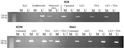
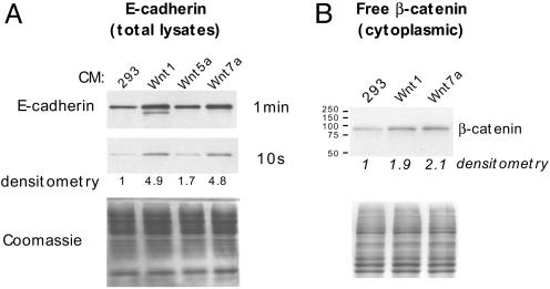
E-cadherin loss in cancer is associated with de-differentiation, invasion, and metastasis. Drosophila DE-cadherin is regulated by Wnt/beta-catenin signaling, although this has not been demonstrated in mammalian cells. We previously reported that expression of WNT7a, encoded on 3p25, was frequently downregulated in lung cancer, and that loss of E-cadherin or beta-catenin was a poor prognostic feature. Here we show that WNT7a both activates E-cadherin expression via a beta-catenin specific mechanism in lung cancer cells and is involved in a positive feedback loop. Li+, a GSK3 beta inhibitor, led to E-cadherin induction in an inositol-independent manner. Similarly, exposure to mWNT7a specifically induced free beta-catenin and E-cadherin. Among known transcriptional suppressors of E-cadherin, ZEB1 was uniquely correlated with E-cadherin loss in lung cancer cell lines, and its inhibition by RNA interference resulted in E-cadherin induction. Pharmacologic reversal of E-cadherin and WNT7a losses was achieved with Li+, histone deacetylase inhibition, or in some cases only with combined inhibitors. Our findings provide support that E-cadherin induction by WNT/beta-catenin signaling is an evolutionarily conserved pathway operative in lung cancer cells, and that loss of WNT7a expression may be important in lung cancer development or progression by its effects on E-cadherin.


Figures

Fig. 1.
Fig. 1.
Induction of E-cadherin by Li+ and TSA in lung tumor cell lines. (A) Cells were exposed to Li+ (20 mM) or TSA (0.33 μM) and equal amounts (10 μg) of protein lysates analyzed by Western blot with anti-E-cadherin antibodies. Equal loading was confirmed by parallel gels stained with Coomassie blue (not shown). (B) Additive effects of combined Li+ (10 mM) and TSA (41 nM) in H661 cells. (C) Myo-inositol does not block E-cadherin induction by Li+. H661 cells were treated with 20 mM Li+ for 24 h with or without 2 mM myo-inositol and analyzed by Western blot. Subsequent tubulin detection confirmed equal loading. (D) Effect of 24 h treatment with Li+ (20 mM), TSA (0.33 μM), or the combination on E-cadherin protein induction in additional lung cancer cell lines as described in the text.
Fig. 2.
Fig. 2.
E-cadherin promoter methylation analysis. E-cadherin-expressing and -negative cell lines were examined for promoter methylation. Only selected examples are shown. Lymphocyte DNA was used as a source of unmethylated DNA, and in vitro methylated DNA was used as a positive control. Cells were untreated, or treated for 24 h with Li+ (20 mM), TSA (0.33 μM), or both. After DNA extraction and sodium bisulfite treatment, PCR was performed for 35 cycles with primers specific for methylated (M) or unmethylated (U) templates, and the products were analyzed on agarose gels.
Fig. 3.
Fig. 3.
Wnt7a induces E-cadherin by canonical WNT/β-catenin signaling. (A) H661 cells were exposed to cultured supernatants of control 293 cells, or 293 cells stably transfected with mWnt1, mWnt5a, or mWnt7a. After 44 h, cells were harvested and 10 μg of protein extracts per lane were analyzed by Western blot for E-cadherin. Equal loading was confirmed by Coomassie blue staining. Densitometry values are relative to the 293 control supernatant set at 1.0. (B) Increased cytoplasmic β-catenin in H661 cells is induced by cultured supernatants from transfected 293 cells expressing mWnt1 and mWnt7a. H661 cells were exposed to cultured supernatants for 6 h. Twenty micrograms of the cytoplasmic fraction was analyzed for β-catenin. A parallel gel was stained with Coomassie blue for loading.

References

    1. Wistuba, I. I., Behrens, C., Virmani, A. K., Mele, G., Milchgrub, S., Girard, L., Fondon, J. W., III, Garner, H. R., McKay, B., Latif, F., et al. (2000) Cancer Res. 60, 1949-1960. - PubMed
    1. Burbee, D. G., Forgacs, E., Zochbauer-Muller, S., Shivakumar, L., Fong, K., Gao, B., Randle, D., Kondo, M., Virmani, A., Bader, S., et al. (2001) J. Natl. Cancer Inst. 93, 691-699. - PMC - PubMed
    1. Tomizawa, Y., Kohno, T., Kondo, H., Otsuka, A., Nishioka, M., Niki, T., Yamada, T., Maeshima, A., Yoshimura, K., Saito, R., et al. (2002) Clin. Cancer Res. 8, 2362-2368. - PubMed
    1. Tomizawa, Y., Sekido, Y., Kondo, M., Gao, B., Yokota, J., Roche, J., Drabkin, H., Lerman, M. I., Gazdar, A. F. & Minna, J. D. (2001) Proc. Natl. Acad. Sci. USA 98, 13954-13959. - PMC - PubMed
    1. Tse, C., Xiang, R. H., Bracht, T. & Naylor, S. L. (2002) Cancer Res. 62, 542-546. - PubMed

Publication types