. 2003 Sep 1;162(5):757-63.
doi: 10.1083/jcb.200305048. Epub 2003 Aug 25.

Interaction of Aurora-A and centrosomin at the microtubule-nucleating site in Drosophila and mammalian cells

Affiliations

Interaction of Aurora-A and centrosomin at the microtubule-nucleating site in Drosophila and mammalian cells

Yasuhiko Terada et al. J Cell Biol. .

Abstract

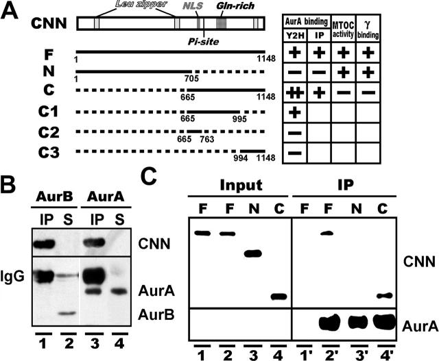
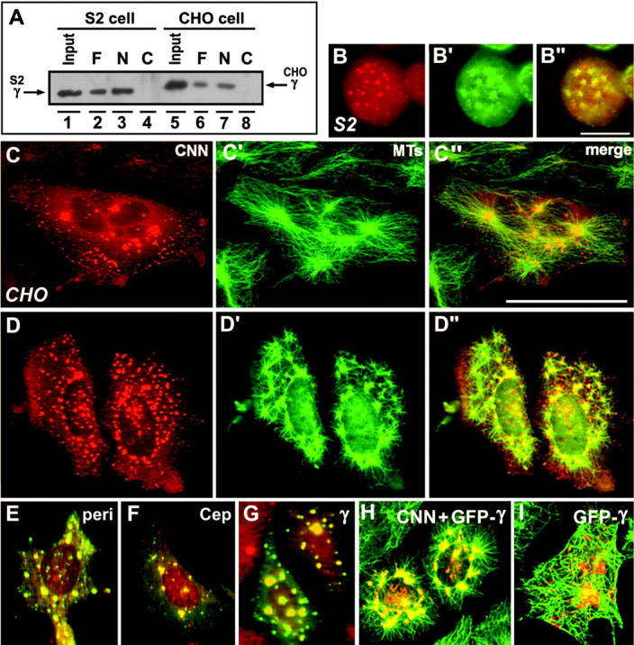
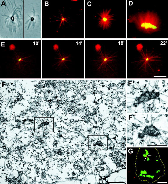
A mitosis-specific Aurora-A kinase has been implicated in microtubule organization and spindle assembly in diverse organisms. However, exactly how Aurora-A controls the microtubule nucleation onto centrosomes is unknown. Here, we show that Aurora-A specifically binds to the COOH-terminal domain of a Drosophila centrosomal protein, centrosomin (CNN), which has been shown to be important for assembly of mitotic spindles and spindle poles. Aurora-A and CNN are mutually dependent for localization at spindle poles, which is required for proper targeting of gamma-tubulin and other centrosomal components to the centrosome. The NH2-terminal half of CNN interacts with gamma-tubulin, and induces cytoplasmic foci that can initiate microtubule nucleation in vivo and in vitro in both Drosophila and mammalian cells. These results suggest that Aurora-A regulates centrosome assembly by controlling the CNN's ability to targeting and/or anchoring gamma-tubulin to the centrosome and organizing microtubule-nucleating sites via its interaction with the COOH-terminal sequence of CNN.


Figures

Figure 1.
Figure 1.
Interaction of Aurora-A with CNN and γ-tubulin. (A) A map of CNN and its deletion constructs capable of association with Aurora-A assayed by yeast two-hybrid screening (Y2H) and immunoprecipitation (IP). Microtubule-nucleating activity (MTOC activity) and γ-tubulin interaction (γ binding) are also summarized on the right. CNN contains characteristic regions, including leucine zipper motifs (Leu zipper), a potential nuclear localization signal (NLS), a putative Aurora-A phosphorylation site (Pi-site), and a glutamine-enriched region (Gln-rich). Numbers indicate the positions of amino acids. (B) HA-tagged CNN binds to Aurora-A (lanes 3 and 4), but not Aurora-B (lanes 1 and 2), in S2 cells. Proteins in immunoprecipitated (IP) and nonprecipitated supernatant (S) fractions, prepared from HA-CNN–expressing (lanes 1 and 3) and –nonexpresssing (lanes 2 and 4) cells, were identified by blotting with HA, Aurora-A, and Aurora-B antibodies. (C) Aurora-A binds to the COOH-terminal domain of CNN. In vitro synthesized full (F), NH2-terminal (N), and COOH-terminal (C) domains of CNN (Input, lanes 1 to 4) were mixed with (IP, lanes 2′ to 4′) and without (IP, lane 1′) His-tagged Aurora-A purified from bacteria.
Figure 2.
Figure 2.
Immunolocalization of centrosome proteins at the spindle pole. (A) Double staining of mitotic S2 cells with Aurora-A/γ-tubulin and CNN/γ-tubulin after depletion of either Aurora-A (a–f) or CNN (g–l) by RNAi. DAPI staining is also shown in cells labeled with γ-tubulin (gamma). Different amounts of Aurora-A and CNN remained at the pole in depleted cells. Aurora-A and CNN are dependent on one another to localize at the spindle pole, and two proteins are required for recruiting γ-tubulin to the centrosome. (B) Localization of CP190 (a′–c′) and CP60 (d′–f′) in control (a and d), Aurora-A–depleted (b and e), and CNN-depleted (c and f) cells. The cells were also stained with α-tubulin and DAPI (a–f). Although depletion of Aurora-A and CNN predominantly induced abnormal spindles in monopolar/multipolar organization, spindles in bipolar orientation were selected to demonstrate the absence of centrosomal proteins at each pole. Bars, 10 μm.
Figure 3.
Figure 3.
CNN interacts with γ-tubulin and induces microtubule-nucleating sites. (A) The NH2-terminal domain of CNN binds to γ-tubulin. Nickel beads conjugated with His-tagged full (F), NH2-terminal (N), and COOH-terminal (C) domains of CNN were incubated with cell extracts prepared from mitotic S2 (lanes 2–4) and CHO cells (lanes 6–8). Endogenous γ-tubulin is shown in lanes 1 and 5. (B–D) Immunostaining of HA-CNN–expressing S2 (B) and CHO cells (C–D) with HA (B–D) and α-tubulin antibodies (B′–D′). Merged images were shown in B′′–D′′. CNN expression caused the formation of cytoplasmic aggregates associated with microtubule asters. In D–D′′, cells were briefly recovered from nocodazole treatment before fixation. (E–G) Colocalization of pericentrin (E), Cep135 (F), and γ-tubulin (G) at the cytoplasmic foci induced by CNN expression. Merged images of CNN (green) and other centrosomal proteins (red) were shown. (H–I) Induction of GFP-tagged γ-tubulin in CHO cells coexpressing (H) or not coexpressing HA-CNN (I). To merge images, GFP was converted to red in double-stained cells with microtubules (green) and HA (green). Although expression of γ-tubulin alone induced cytoplasmic dots, microtubule-nucleating activity of the aggregates was detected only when γ-tubulin was coexpressed with CNN. Bars, 10 μm (B) and 50 μm (C–I).
Figure 4.
Figure 4.
Cytoplasmic aggregates induced by CNN overexpression in mammalian cells. (A–D) In vitro microtubule nucleation onto CNN-containing sites detected by phase-contrast (A) and fluorescence (B–D) microscopy. GFP-tagged CNN aggregates were fractionated from CHO cells and incubated with X-rhodamine–conjugated brain tubulin. CNN aggregates in different sizes and shapes nucleated various numbers of microtubules. (E) Time-lapse images of GFP-tagged CNN-containing aggregates with assembled microtubules. CNN aggregates mixed with X-rhodamine tubulin were placed on a microscopic stage at time zero, and fluorescence images were taken at indicated times after the temperature was shifted to 37°C. (F) Thin-section EM of CHO cells expressing GFP-tagged CNN. The cells were briefly extracted with a detergent containing microtubule-stabilizing buffer before fixation. Two microtubule asters are seen in the field, and there is an electron-dense particle of different shape at each center. F′ and F′′ are close-ups of the areas outlined in F. (G) Immunofluorescence staining of 293 cells with anti-human centrin-2 antibodies (red). Dotted lines indicate the outline of a cell expressing CNN aggregates (green). There are two centrioles (arrows) that were not included in all sites induced by CNN overexpression. Bars, 10 μm (A–E, and G) and 1 μm (F–F′′).

References

    1. Barbosa, V., R.R. Yamamoto, D.S. Henderson, and D.M. Glover. 2000. Mutation of a Drosophila gamma tubulin ring complex subunit encoded by discs degenerate-4 differentially disrupts centrosomal protein localization. Genes Dev. 14:3126–3139. - PMC - PubMed
    1. Berdnik, D., and J.A. Knoblich. 2002. Drosophila Aurora-A is required for centrosome maturation and actin-dependent asymmetric protein localization during mitosis. Curr. Biol. 12:640–647. - PubMed
    1. Bischoff, J.R., L. Anderson, Y. Zhu, K. Mossie, L. Ng, B. Souza, B. Schryver, P. Flanagan, F. Clairvoyant, C. Ginthe, et al. 1998. A homologue of Drosophila aurora kinase is oncogenic and amplified in human colorectal cancers. EMBO J. 17:3052–3065. - PMC - PubMed
    1. Eyers, P.A., E. Erikson, L.G. Chen, and J.L. Maller. 2003. A novel mechanism for activation of the protein kinase Aurora A. Curr. Biol. 13:691–697. - PubMed
    1. Giet, R., D. McLean, S. Descamps, M.J. Lee, J.W. Raff, C. Prigent, and D.M. Glover. 2002. Drosophila Aurora A kinase is required to localize D-TACC to centrosome and to regulate astral microtubules. J. Cell Biol. 156:437–451. - PMC - PubMed

Publication types