. 2003 Sep 1;198(5):725-36.
doi: 10.1084/jem.20021098. Epub 2003 Aug 25.

Stroke-induced immunodeficiency promotes spontaneous bacterial infections and is mediated by sympathetic activation reversal by poststroke T helper cell type 1-like immunostimulation

Affiliations

Stroke-induced immunodeficiency promotes spontaneous bacterial infections and is mediated by sympathetic activation reversal by poststroke T helper cell type 1-like immunostimulation

Konstantin Prass et al. J Exp Med. .

Abstract

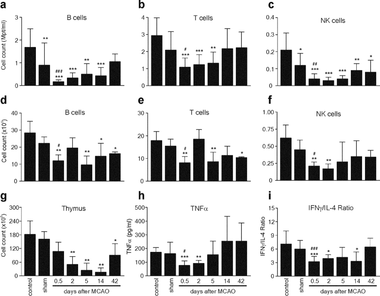
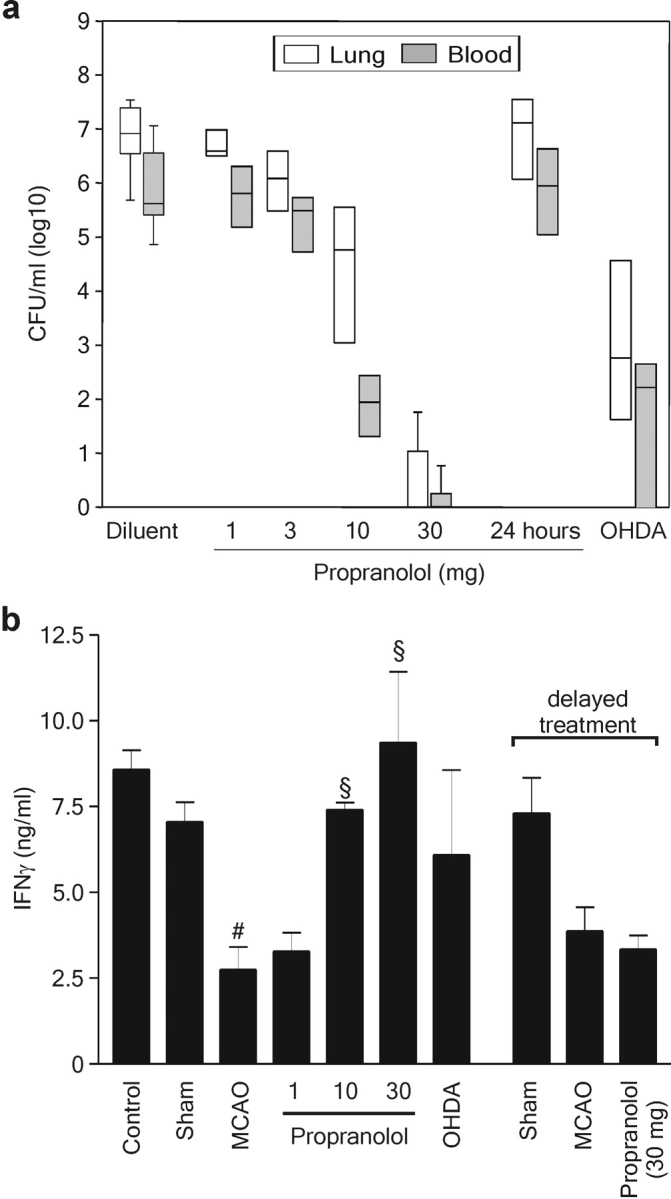
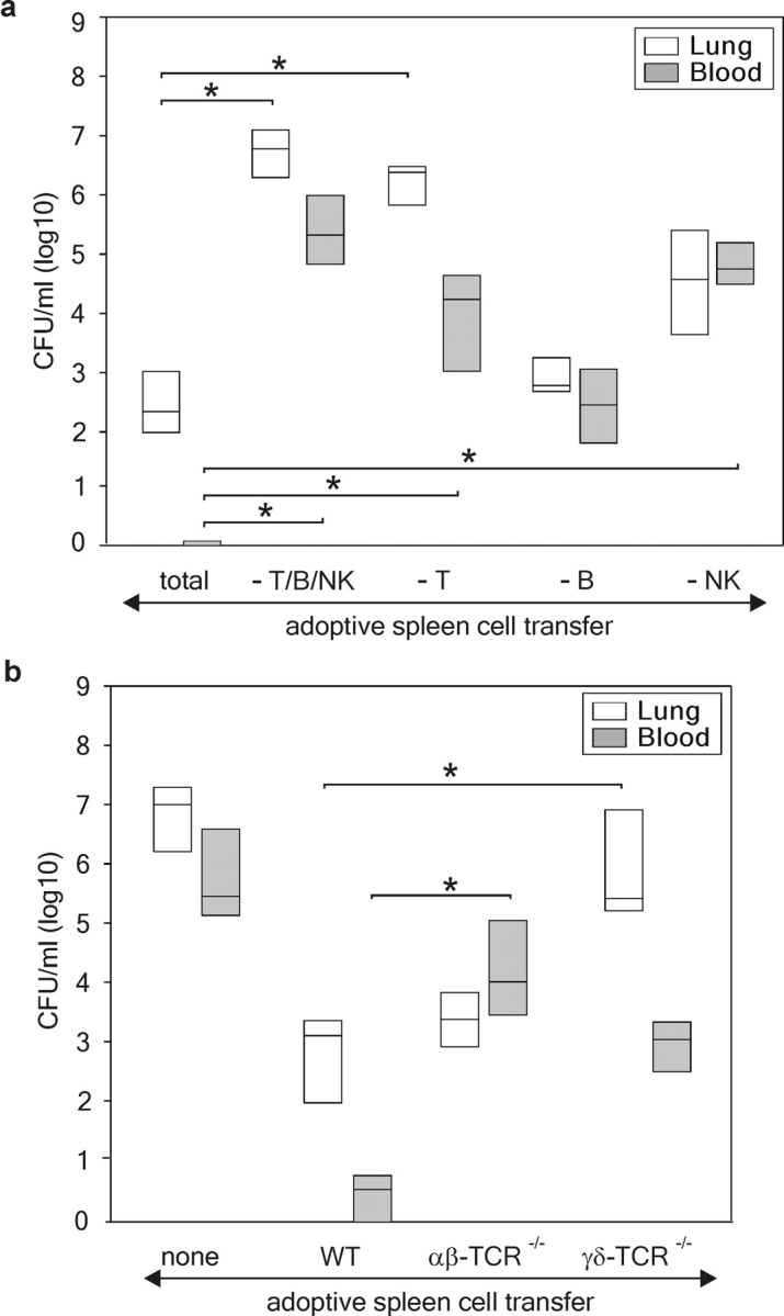
Infections are a leading cause of death in stroke patients. In a mouse model of focal cerebral ischemia, we tested the hypothesis that a stroke-induced immunodeficiency increases the susceptibility to bacterial infections. 3 d after ischemia, all animals developed spontaneous septicemia and pneumonia. Stroke induced an extensive apoptotic loss of lymphocytes and a shift from T helper cell (Th)1 to Th2 cytokine production. Adoptive transfer of T and natural killer cells from wild-type mice, but not from interferon (IFN)-gamma-deficient mice, or administration of IFN-gamma at day 1 after stroke greatly decreased the bacterial burden. Importantly, the defective IFN-gamma response and the occurrence of bacterial infections were prevented by blocking the sympathetic nervous system but not the hypothalamo-pituitary-adrenal axis. Furthermore, administration of the beta-adrenoreceptor blocker propranolol drastically reduced mortality after stroke. These data suggest that a catecholamine-mediated defect in early lymphocyte activation is the key factor in the impaired antibacterial immune response after stroke.


Figures

Figure 1.
Figure 1.
Stroke induces bacteremia and pneumonia. (a) Lungs and blood samples from sham (n = 10) and MCAO-treated mice (n = 4–14 per group) were collected for bacteriological analysis at the indicated time points after surgery. Data are given in CFU/ml (log 10) blood or lung tissue homogenate. (b) In a different set of experiments, lungs from sham-operated (n = 4) and MCAO mice (n = 4) were collected after 72 h for histological examination. A representative 12-μm section of HE-stained lung from MCAO but not from sham animals revealed signs (thickening of alveolar walls and neutrophilic infiltrates) of E. coli pneumonia. ×160.
Figure 2.
Figure 2.
Stroke induces long-lasting lymphopenia and impaired cytokine expression. Spleen, thymus, and peripheral blood samples of untreated SV129/J mice (control) and sham or MCAO mice were collected at different time points (sham, 12 h; MCAO, as indicated) after surgery. Leukocyte counts in blood (a–c), spleen (d–f), and thymus (g) single cell suspensions were determined as described in Materials and Methods. The lymphocyte subsets were determined by flow cytometry and their absolute numbers were calculated. (h and i) Aliquots of blood samples were stimulated ex vivo with either LPS for analysis of monocytic TNF-α expression or Con A for analysis of IFN-γ and IL-4 synthesis, as described in Materials and Methods. Cytokines were determined in supernatants by ELISA. The Con A–induced lymphokine expression is given as the ratio of IFN-γ and IL-4 production calculated for each individual. Data are shown as mean ± SD. *, results differed from the control group; *, P < 0.05; **, P < 0.01; ***, P < 0.001; #, results differed from the sham-operated group; #, P < 0.05; ##, P < 0.01. Mann-Whitney U test, n = 3–11 per group.
Figure 3.
Figure 3.
Stroke induces increased apoptosis in lymphoid organs. Spleen and thymus from untreated SV129/J mice (control) and sham or MCAO mice were collected 12 h after surgery. Splenocytes (a) and thymocytes (b) were isolated and investigated for apoptosis by annexin V labeling and flow cytometry. Lymphocyte subpopulations were determined by staining with mAbs against cell-type–specific surface markers. Data are shown as mean ± SD. *, results differed from the control group; *, P < 0.05; **, P < 0.01; ***, P < 0.001; #, results differed from the sham-operated group; #, P < 0.05; ##, P < 0.01; ###, P < 0.001. Mann-Whitney U test, n = 8 per group. (c) Thymic tissue sections from sham- and MCAO-treated mice were stained with HE (top) or examined by the fluorescent TUNEL method (bottom). Thymi from MCAO mice showed many apoptotic cells with typical features of nuclear condensation and fragmentation (HE; ×400). Note also the high number of TUNEL+ cells (bright red nuclei) in the thymus cortex from MCAO animals as compared with sham mice. ×100.
Figure 4.
Figure 4.
Effects of β-adrenoreceptor and glucocorticoid receptor blockade on stroke-induced immunodepression and bacterial infection. SV129/J mice underwent sham or MCAO surgery (n = 9–12 per group). MCAO mice received the β-adrenoreceptor antagonist propranolol, the glucocorticoid receptor inhibitor RU486, or only diluent before or after surgery, as described in Materials and Methods. Spleens and blood samples were collected 12 h after surgery. (a) Splenocytes were isolated and investigated for apoptosis by annexin V labeling, staining with mAbs against cell-type–specific surface markers, and flow cytometry. (b) Total leukocyte counts in blood samples were determined as described in Materials and Methods. The percentages of different lymphocyte classes were determined by flow cytometry using mAbs against cell-type–specific surface markers and their absolute numbers calculated. (c) For determination of cytokine production, blood samples were processed and cytokine expression was analyzed as described in Fig. 2. Data are shown as mean ± SD. #, results differed from the sham-operated group; #, P < 0.05; ##, P < 0.01; §, results differed from the diluent-treated MCAO group; §, P < 0.05; §§, P < 0.01; §§§, P < 0.001. Mann-Whitney U test, n = 9–12 per group. (d) In a different set of experiments, lungs and blood samples from sham and inhibitor- or diluent-treated MCAO mice were collected for bacteriological analysis 72 h after surgery. Data are shown as box plots in CFU/ml (log 10) blood or lung tissue homogenate.
Figure 5.
Figure 5.
Prevention of bacterial infections and restoration of defective IFN-γ response by SNS inhibitors is dose and time dependent. (a) MCAO mice were treated with diluent or different doses of propranolol (3 × 1–3 × 30 mg/kg BW) commencing immediately after MCAO or 24 h (3 × 30 mg/kg BW) after MCAO, or with 6-OHDA before MCAO as described in Materials and Methods and as indicated (n = 4 in each group). Lungs and blood samples were collected for bacteriological analysis 72 h after MCAO. Data are shown as box plots in CFU/ml (log 10) blood or lung tissue homogenate. (b) In a different experiment, spleens of untreated mice (control), sham-, or MCAO-operated mice, which received diluent, propranolol, or 6-OHDA as described above, were collected 12 or 36 h (delayed propranolol treatment) after surgery. 106/ml spleen cells were stimulated ex vivo with Con A for 24 h and IFN-γ production was analyzed by ELISA, as described in Materials and Methods. Data are shown as mean ± SD. #, results differed from the sham-operated group; #, P < 0.05; §, results differed from the MCAO animals; §, P < 0.05. Mann-Whitney U test, n = 3–6 per group.
Figure 6.
Figure 6.
Blocking the SNS improves survival after experimental stroke. Mice underwent MCAO and received either diluent or 10 mg/kg BW immediately before, 4, and 8 h after MCAO (n = 15 per group). Differences in survival between propranolol-treated versus diluent-treated mice were tested for significance on day 11 after MCAO and are indicated by asterisks. *, P < 0.05. Fisher's exact test.
Figure 7.
Figure 7.
Adoptive lymphocyte transfer after experimental stroke prevents infection. (a) Adoptive lymphocyte transfer was performed 24 h after MCAO using either unseparated (total) or T, B, and NK cell, or T cell–, B cell–, or NK cell–depleted spleen cell suspensions from wild-type SV129/J mice. *, results differed from unseparated splenocyte transfer group. Kruskal-Wallis analysis of variance (ANOVA) after pairwise comparison with Dunn's method test, n = 4 per group. (b) In a different set of experiments, splenocytes from αβ T cell–deficient (αβ-TCR−/−), γδ T cell–deficient (γδ-TCR−/−), or wild-type B6 mice (WT) were transferred into B6 mice 24 h after MCAO. 48 h later, blood and lung samples were analyzed for bacterial counts. Data are shown as box plots in CFU/ml (log 10) blood or lung tissue homogenate. *, results differed from WT splenocyte transfer group. Kruskal-Wallis analysis of variance (ANOVA) after comparison of pairs with Dunn's method test, n = 8 per group.
Figure 8.
Figure 8.
IFN-γ is essential in preventing the bacterial infections. (a) Adoptive lymphocyte transfer experiments 24 h after MCAO revealed an impaired ability of splenocytes from IFN-γ–deficient mice (IFN-γ−/−) to prevent bacterial infection in blood and lung when compared with splenocytes from wild-type B6 mice (n = 8 per group). (b) Administration of 2 μg recombinant IFN-γ 24 h, but not 48 h, after cerebral ischemia reduced the bacterial burden in peripheral blood and lungs as measured 72 h after MCAO (n = 4 per group). For data representation see Fig. 5.

Comment in

  • Effects of stroke beyond the brain.
    Wong CHY. Wong CHY. Nat Rev Immunol. 2019 Dec;19(12):719. doi: 10.1038/s41577-019-0234-4. Nat Rev Immunol. 2019. PMID: 31624320 No abstract available.

Similar articles

Cited by

References

    1. Davenport, R.J., M.S. Dennis, I. Wellwood, and C.P. Warlow. 1996. Complications after acute stroke. Stroke. 27:415–420. - PubMed
    1. Johnston, K.C., J.Y. Li, P.D. Lyden, S.K. Hanson, T.E. Feasby, R.J. Adams, R.E. Faught, Jr., and E.C. Haley, Jr. 1998. Medical and neurological complications of ischemic stroke: experience from the RANTTAS trial. RANTTAS investigators. Stroke. 29:447–453. - PubMed
    1. Georgilis, K., A. Plomaritoglou, U. Dafni, Y. Bassiakos, and K. Vemmos. 1999. Aetiology of fever in patients with acute stroke. J. Intern. Med. 246:203–209. - PubMed
    1. Grau, A.J., F. Buggle, P. Schnitzler, M. Spiel, C. Lichy, and W. Hacke. 1999. Fever and infection early after ischemic stroke. J. Neurol. Sci. 171:115–120. - PubMed
    1. Langhorne, P., D.J. Stott, L. Robertson, J. MacDonald, L. Jones, C. McAlpine, F. Dick, G.S. Taylor, and G. Murray. 2000. Medical complications after stroke: a multicenter study. Stroke. 31:1223–1229. - PubMed

Publication types

MeSH terms