

Comparison of memory fMRI response among normal, MCI, and Alzheimer's patients
- PMID: 12939424
- PMCID: PMC2744465
- DOI: 10.1212/01.wnl.0000079052.01016.78
Comparison of memory fMRI response among normal, MCI, and Alzheimer's patients
Erratum in
- Neurology. 2003 Oct 28;61(8):1164
Abstract
Objective: To determine whether an fMRI memory encoding task distinguishes among cognitively normal elderly individuals, patients with mild cognitive impairment (MCI), and patients with early Alzheimer's disease (AD).
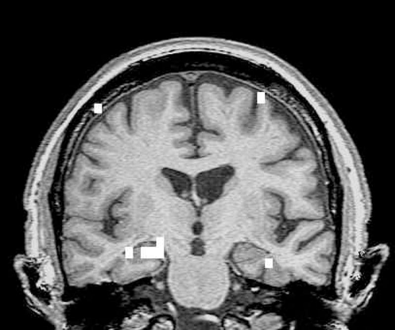
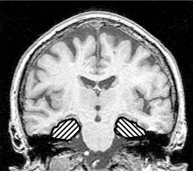
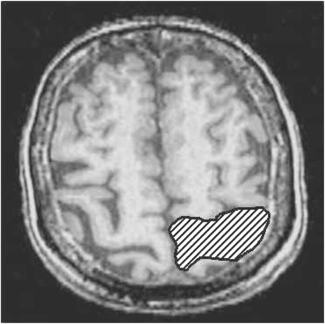
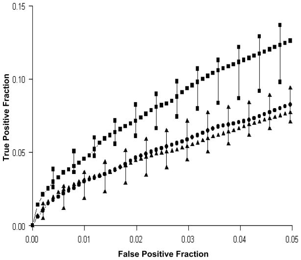
Methods: Twenty-nine subjects (11 normal, 9 MCI, 9 AD) were studied with an fMRI memory encoding task. A passive sensory task was also performed to assess potential intergroup differences in fMRI responsiveness. Activation in the medial temporal lobe for the memory task and in the anatomic rolandic area for the sensory task was studied. Intergroup comparisons were performed using receiver operating characteristic (ROC) analyses. The ROC method provides rigorous control of artifactual false-positive "activation." Subjects were tested for recall and recognition of the encoding task stimuli following the fMRI study.
Results: Medial temporal lobe activation was greater in normal subjects than MCI and AD patients (p = 0.03 and p = 0.04). There was no difference between AD and MCI patients in fMRI memory performance [corrected]. There was an association between fMRI memory activation (area under the ROC curve) and post-fMRI performance on recognition and free recall. There was no difference among the three groups on the sensory task.
Conclusions: MCI and AD patients had less medial temporal lobe activation on the memory task than the normal subjects but similar activation as normal subjects on the sensory task. These findings suggest decreased medial temporal activation may be a specific marker of limbic dysfunction due to the neurodegenerative changes of AD. In addition, fMRI is sufficiently sensitive to detect changes in the prodromal, MCI, phase of the disease.
Figures
References
-
- Buckner R, Snyder A, Sanders A, Raichle M, Morris J. Functional brain imaging of young, nondemented, and demented older adults. Journal of Cognitive Neuroscience. 2000;12:24–34. - PubMed
-
- Johnson S, Saykin A, Baxter L, et al. The relationship between fMRI activation and cerebral atrophy: comparison of normal aging and Alzheimer disease. NeuroImage. 2000;11:179–187. - PubMed
-
- Prvulovic D, Hubl D, Sack A, et al. Functional imaging of visuospatial processing in Alzheimer’s disease. NeuroImage. 2002;17:1403–1414. - PubMed
Publication types
MeSH terms
Grants and funding
LinkOut - more resources
Full Text Sources
Other Literature Sources
Medical