. 2003 Sep 1;22(17):4375-84.
doi: 10.1093/emboj/cdg418.

Binding, activation and dissociation of the dimeric SecA ATPase at the dimeric SecYEG translocase

Affiliations

Binding, activation and dissociation of the dimeric SecA ATPase at the dimeric SecYEG translocase

Franck Duong. EMBO J. .

Abstract

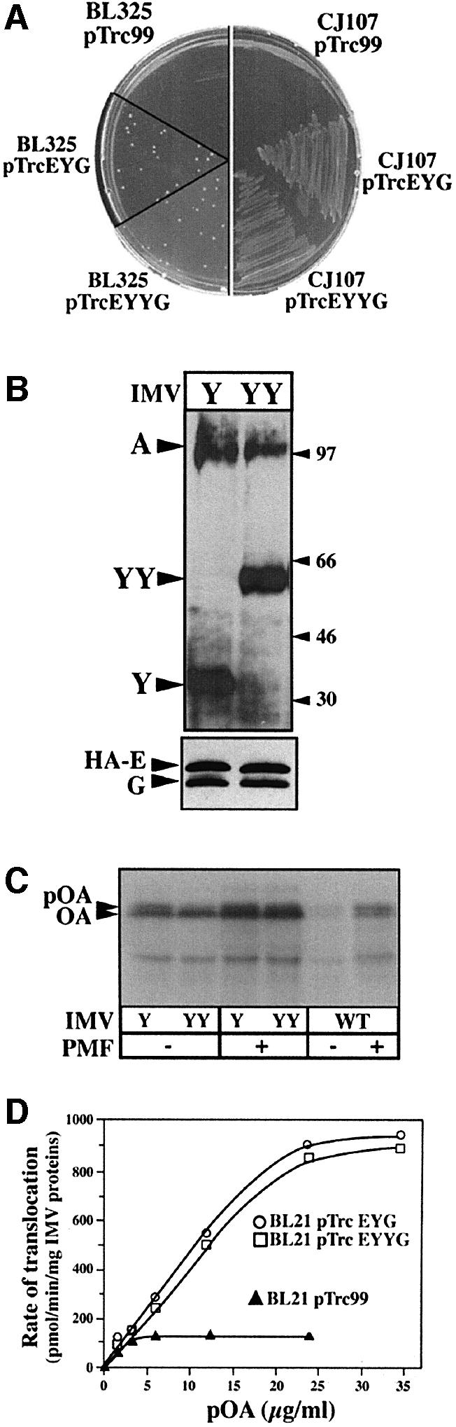
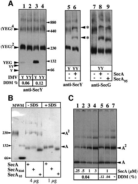
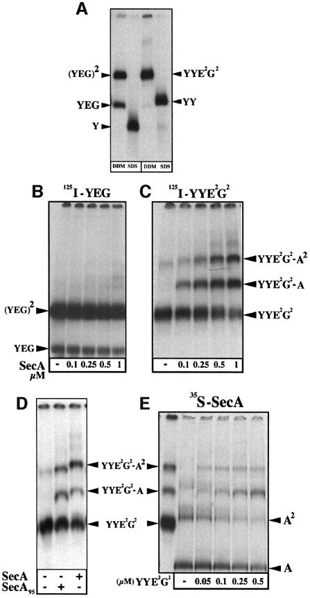
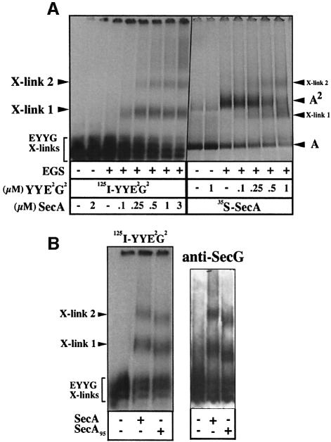
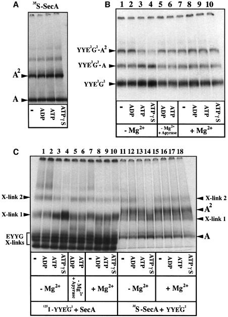
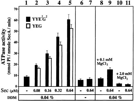
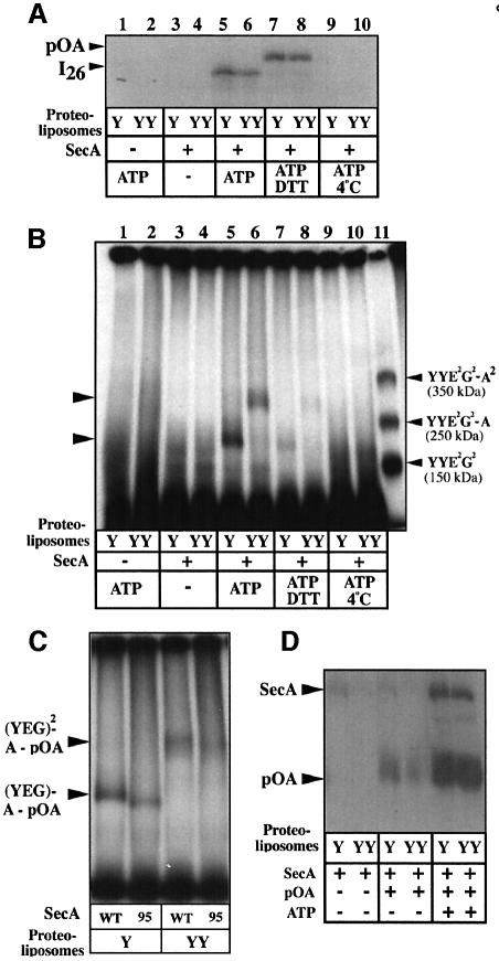
The bacterial preprotein translocase is comprised of a membrane-embedded oligomeric SecYEG structure and a cytosolic dimeric SecA ATPase. The associations within SecYEG oligomers and SecA dimers, as well as between these two domains are dynamic and reversible. Here, it is shown that a covalently linked SecYEG dimer forms a functional translocase and a high affinity binding site for monomeric and dimeric SecA in solution. The interaction between these two domains stimulates the SecA ATPase, and nucleotides modulate the affinity and ratio of SecA monomers and dimers bound to the linked SecYEG complex. During the translocation reaction, the SecA monomer remains in stable association with a SecYEG protomer and the translocating preprotein. The nucleotides and translocation-dependent changes of SecA-SecYEG associations and the SecA dimeric state may reflect important facets of the preprotein translocation reaction.


Figures

None
Fig. 1. The covalently linked SecYEG dimer is functional for pre protein translocation. (A) Overproduction of SecYYEG suppresses the SecDFyajC cold-sensitive and SecY thermo-sensitive phenotypes. Strain BL325 (BL21 tgt::kan-araC+ -PBAD::yajCsecDF) or CJ107 (Wolfe et al., 1985) were transformed with the isopropyl-β-d-thio galactopyranoside (IPTG)-inducible plasmid pTrc99 or pTrc99 encoding SecYEHAG (pTrcEYG) or SecYYEHAG (pTrcEYYG). Transformants were plated on LB/ampicillin (50 µg/ml) contain ing 1 mM IPTG and incubated at the restrictive temperature. (B) Immunostaining of IMV preparations. About 1 µg of IMV proteins prepared from E.coli BL21 overexpressing SecYEHAG (IMVs-Y) or SecYYEHAG (IMVs-YY) were analyzed by SDS–PAGE and immunostained with the indicated antibodies. In the presence of SDS, SecY (48.5 kDa) and SecYY (97 kDa) migrate aberrantly. (C and D) Translocase activity of the IMV preparations. Translocation reactions were performed and analyzed as described in Materials and methods.
None
Fig. 2. Oligomeric behavior of covalently-linked SecYEG dimer and SecA. (A) Immunodetection of SecYEG oligomers in membranes enriched with SecYEG (IMVs-Y) or SecYYE2G2 (IMVs-YY). IMVs were solubilized in TSG buffer at the indicated detergent concentration. Protein aliquots (∼0.2 µg) were analyzed by linear gradient BN–PAGE (4–12%) and immunostained. Where indicated, IMVs were incubated with SecA or SecA95 (4 µg; 0.8 µM protomer). The asterisks indicate the probable complex of SecYYE2G2 with monomeric and dimeric SecA. (B) SecA, His6-tagged SecA or His6-tagged SecA95 in TSG buffer (±SDS) were analyzed on a native gel followed by Coomassie blue staining. Molecular weight markers are BSA and catalase. (C) [35S]SecA (∼20 000 c.p.m., ∼100 ng) was mixed at room temperature with unlabeled SecA to reach the desired concentration. Samples were diluted in TSG buffer containing DDM and analyzed by BN–PAGE (4–10%) and autoradiography. The final SecA and DDM concentrations in each sample are indicated.
None
Fig. 3. The stabilized SecYEG dimer forms a stable binding site for SecA monomers and dimers. (A) Oligomeric state of the purified SecYEG complexes. [125I]SecYEG and [125I]SecYYE2G2 purified complexes (∼30 000 c.p.m., ∼20 ng, in TSG buffer 0.03% DDM or 0.1% SDS) were analyzed by BN–PAGE (5–13%) and autoradio graphy. (BD) Complexes between radiolabeled SecYEG and unlabeled SecA. [125I]SecYEG and [125I]SecYYE2G2 purified complexes (∼30 000 c.p.m., 0.7 µM) were incubated (5 min, 22°C) with SecA or SecA95 at the indicated concentration in TSG buffer containing 0.04% DDM. The resulting complexes were separated by BN–PAGE (4–12%) and revealed by autoradiography. (E) Complexes between radiolabeled SecA and unlabeled SecYYE2G2. [35S]SecA (∼20 000 c.p.m., 1 µM) was incubated in TSG buffer containing 0.04% DDM with the indicated concentration of the purified SecYYE2G2 complex. The resulting complexes were analyzed by BN–PAGE (4–12%) and autoradiography. For Mr reference, a sample of the [125I]SecYYE2G2 complex incubated with unlabeled SecA is loaded in the left lane of the gel. Calculated molecular weights of the Sec proteins and Sec complexes: SecY (48.5 kDa); SecYY (97 kDa); His-SecE (14 kDa); SecG (11 kDa); His-SecA (103 kDa); His-SecA95 (95 kDa); SecYEG (73.5 kDa); SecY2E2G2 (147 kDa); SecY4E4G4 (294 kDa); SecY2E2G2–SecA (250 kDa); SecY2E2G2–SecA2 (353 kDa).
None
Fig. 4. Cross-linking production between SecYYE2G2 and SecA. (A) [125I]SecYYE2G2 (0.1 µM) or [35S]SecA (1 µM) were incubated in CL buffer (5 min, 22°C) with the indicated concentration of unlabeled SecYYE2G2 or unlabeled SecA, respectively. After cross-linking by EGS and dissociation of the non-covalent protein complexes by SDS, samples were analyzed by BN–PAGE (4–12%) and autoradiography. (B) [125I]SecYYE2G2 or unlabeled SecYYE2G2 were incubated as in (A) with SecA or SecA95, and cross-links were revealed by autoradiography or immunostaining.
None
Fig. 5. Nucleotide-dependent SecA–SecYEG associations. (A) Nucleotides do not change the oligomeric state of SecA in solution. [35S]SecA (∼20 000 c.p.m., 1 µM) was incubated (10 min, 22°C) in TSG buffer containing 0.04% DDM with the indicated nucleotides (1 mM). Samples were analyzed by BN–PAGE (4–12%) and autoradiography. (B) Complexes between radiolabeled SecYEG and unlabeled SecA after addition of nucleotides. [125I]SecYYE2G2 (0.7 µM) was mixed with SecA (1 µM) in TSG buffer (0.04% DDM; ±5 mM MgCl2). After incubation (5 min, 22°C), nucleotides were added and the incubation was prolonged for 5 min. Samples (lanes 5 and 6) were treated with potato apyrase (grade VII; 20 U/ml) for an additional 2 min. All samples were returned to ice before analysis by BN–PAGE (4–12%) and autoradiography. (C) Cross-link production between SecYYE2G2 and SecA after addition of nucleotides. [125I]SecYYE2G2 (0.1 µM) or [35S]SecA (1 µM) were incubated in CL buffer (±5 mM MgCl2) with unlabeleled SecA (2 µM) or unlabeled SecYYE2G2 (0.5 µM), respectively. After incubation with nucleotides (as described in B), samples were treated by EGS, then dissolved by SDS and analyzed by BN–PAGE (4–12%) and autoradiography.
None
Fig. 6. Activation of the SecA ATPase by SecYEG. SecA (0.8 µM) was incubated at room temperature with the purified SecYEG or SecYYE2G2 complexes in TSG buffer (±0.04% DDM; ±MgCl2) containing 2 mM of ATP. The Sec concentration refers to that of the SecYEG protomer (SecYYE2G2 = 2 × SecYEG). After 10 min incubation at 22°C, the inorganic phosphate produced by the SecA ATPase was measured by the photocolorimetric method of Lanzetta et al. (1979).
None
Fig. 7. The SecA monomer is engaged with preprotein and the SecYEG channel during translocation. (A) [125I]ProOmpA–BPTI (∼100 000 c.p.m.; 4 µg/ml) was incubated (10 min, 37°C) in TL buffer (50 µl final volume) with SecYEG- or SecYYE2G2-proteoliposomes (15 µg/ml), BSA (200 µg/ml), SecA (80 µg/ml; 0.8 µM) and ATP (2 mM) or DTT (2 mM) where indicated. Reactions were terminated by addition of apyrase (20 U/ml). Aliquots of the translocation reactions (1/10 vol.) were treated with proteinase K and analyzed by 12% SDS–PAGE and autoradiography. (B) The remainder of the translocation reaction performed in (A) was solubilized with an equal volume of TSG buffer containing 0.12% DDM. Aliquots were analyzed by BN–PAGE (4–10%) and autoradiography. For Mr reference, [125I]SecYYE2G2 complexed to SecA was loaded on the same gel (lane 11). (C) As in (B) lanes 5 and 6, but using the truncated SecA95 or wild-type SecA to drive translocation in SecYEG- or SecYYE2G2-proteoliposomes. (D) The translocation reactions described in (A) were supplemented with [125I]SecA (∼100 000 c.p.m.; 4 nM). At the end of the incubation (10 min, 37°C), proteoliposomes were solubilized with 300 µl of TSG buffer, 0.1% DDM (10 min, 4°C). After centrifugation (10 min, 100 000 g), the membrane extract was incubated with anti-SecG IgG coupled to protein A–Sepharose beads. Immuno precipitates were washed (3 × 0.3 ml of TSG buffer) and analyzed by 10% SDS–PAGE and autoradiography. Theoretical molecular weight of the SecYEG–SecA–pOA complexes: pOA (37 kDa); SecYEG– pOA (112 kDa); (SecYEG)2–pOA (187 kDa); SecYEG–SecA–pOA (212 kDa); (SecYEG)2–SecA (250 kDa); (SecYEG)2–SecA–pOA (287 kDa); (SecYEG)4–pOA (343 kDa).

Similar articles

Cited by

References

    1. Benach J. et al. (2003) Phospholipid-induced monomerization and signal-peptide-induced oligomerization of SecA. J. Biol. Chem. 278, 3628–3638. - PubMed
    1. Bessonneau P., Besson,V., Collinson,I. and Duong,F. (2002) The SecYEG preprotein translocation channel is a conformationally dynamic and dimeric structure. EMBO J., 21, 995–1003. - PMC - PubMed
    1. Breukink E., Nouwen,N., van Raalte,A., Mizushima,S., Tommassen,J. and de Kruijff,B. (1995) The C terminus of SecA is involved in both lipid binding and SecB binding. J. Biol. Chem., 270, 7902–7907. - PubMed
    1. Breyton C., Haase,W., Rapoport,T.A., Kuhlbrandt,W. and Collinson,I. (2002) Three-dimensional structure of the bacterial protein-translocation complex SecYEG. Nature, 418, 662–665. - PubMed
    1. Cao T.B. and Saier,M.H. (2003) The general protein secretory pathway: phylogenetic analyses leading to evolutionary conclusions. Biochim. Biophys Acta, 1609, 115–125. - PubMed

Publication types

MeSH terms

LinkOut - more resources