. 2003 Sep 1;22(17):4409-20.
doi: 10.1093/emboj/cdg424.

Prickle and Strabismus form a functional complex to generate a correct axis during planar cell polarity signaling

Affiliations

Prickle and Strabismus form a functional complex to generate a correct axis during planar cell polarity signaling

Andreas Jenny et al. EMBO J. .

Abstract

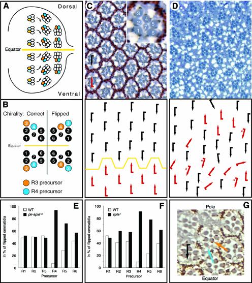
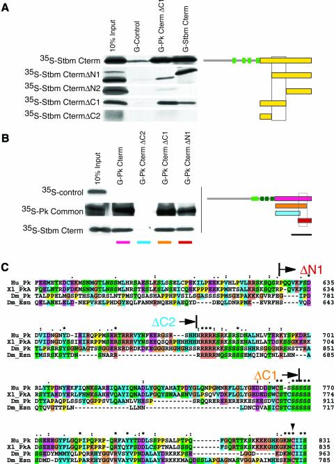
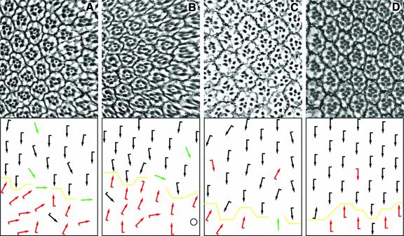
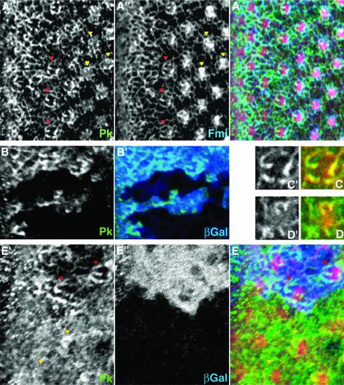
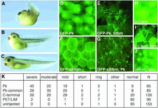
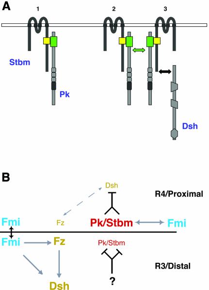
Frizzled (Fz) signaling regulates the establishment of planar cell polarity (PCP). The PCP genes prickle (pk) and strabismus (stbm) are thought to antagonize Fz signaling. We show that they act in the same cell, R4, adjacent to that in which the Fz/PCP pathway is required in the Drosophila eye. We demonstrate that Stbm and Pk interact physically and that Stbm recruits Pk to the cell membrane. Through this interaction, Pk affects Stbm membrane localization and can cause clustering of Stbm. Pk is also known to interact with Dsh and is thought to antagonize Dsh by affecting its membrane localization. Thus our data suggest that the Stbm/Pk complex modulates Fz/Dsh activity, resulting in a symmetry-breaking step during polarity signaling.


Figures

None
Fig. 1. pk is required in the R4 precursor cell. Anterior is left and dorsal is up. Schematic representation of ommatidial patterning in (A) third instar eye disk and (B) adult eye. Clusters rotate in opposing directions with respect to the equator (yellow line) in the eye disk (A) and acquire a dorsal or ventral chirality, as apparent in adult ommatidia (B). R3 precursors are in orange and R4 precursors in blue in both panels. In wild type, the equatorial R3 precursor becomes the anterior tip cell of the cluster, while the polar R4 precursor becomes the posterior recessed cell. Note that in the case of a chirality switch [flipped ommatidia in (B)], the R4 precursor (blue) becomes the R3 cell at the tip of the ommatidium (orange). Tangential sections of adult eyes with a schematic representation underneath are shown in (C) and (D). The insert in (C) shows an enlarged single ommatidium with numbered photoreceptors. (C) Wild-type eye with dorsal (black) and ventral (red arrows) chiral forms separated by equator (yellow line). (D) In pknull (pkpk–sple13), chirality is random (45 ± 3%:53 ± 1%;) and no equator is visible. Thirteen percent of all ommatidia also show rotation defects. Clonal analysis of (Epknull and (Fpksple1 shows that wild-type R4 precursor cells are highly under-represented in ommatidia with wrong chirality: 267 mosaic ommatidia for pknull (54 of which had the wrong chirality) and 416 mosaic ommatidia for pksple1 (60 of which were flipped) were scored. Statistical analysis showed that the apparent under-representation of wild-type R5 precursors is not significant and is due to its proximity to R4 (see text). Thus pk activity is required only in R4. (G) The PkSple isoform present in the R4 precursor only is sufficient for correct choice of chirality. All R3/R4 mosaic ommatidia with the PkSple-expressing construct (marked autonomously by pigment granules) in R4 were wild type. In contrast, those with flipped chirality had PkSple present in the R3 precursor (orange arrow) but not in the R4 precursor (blue arrow; because of the cell fate switch, the R4 precursor becomes R3 and takes the tip position of the ommatidium). The black arrow shows a rescued ommatidium with correct dorsal chirality.
None
Fig. 2. strabismus and prickle dominantly enhance each other’s GOF phenotype. The percentage of ommatidia with planar polarity defects and the standard deviation of sev-Gal4>UAS-PkSple at 25°C and sev-Gal4>UAS-Stbm at 29°C is shown in the first bar (/+) of panels (A) and (B), respectively. The additional bars show the effects of the removal of one copy of the respective gene indicated. The overexpression phenotype of the Sple isoform is dominantly enhanced by the removal of a copy of stbm, while the overexpression phenotype of Stbm is dominantly enhanced by the removal of a copy of pk. Both phenotypes are enhanced by removing a copy of fmi and dgo, but are unaffected by N (see also Table I).
None
Fig. 3. Pk and Stbm proteins interact with each other in vitro. (A) GST fusion proteins with a fragment of the C-terminus of Pk [G-Pk CtermΔC1; orange in scheme in (B)], with the C-terminus of Stbm (G-Stbm Cterm) and with the PkM isoform specific N-terminal region (G-Control) were tested for their ability to pull down the in vitro translated fragments of the C-terminal region of Stbm indicated on left (marked in yellow in the scheme on the right). A fragment of 85 amino acids of the C-terminal cytoplasmic region of Stbm is required for the interaction with Pk and Stbm itself (underlaid with a box in the scheme on the right). As standard, 10% of the in vitro translated protein used for the pull down was loaded directly (10% input). The green boxes in the scheme indicate the four transmembrane domains. (B) Mapping of the binding site of Pk to Stbm and to itself. The GST fusion proteins indicated on top (color coded below, compare with scheme on right) were used to pull down the in vitro translated Stbm Cterm and the in vitro translated region common to all isoforms of Pk. A 131 amino acid fragment of Pk is required for the interaction of Pk with itself and with Stbm (underlaid with a box in scheme on right). The PkM isoform specific N-terminal region (35S-Control) is not interacting with these GST fusion proteins. As standard, 10% of the in vitro translated protein was loaded directly (10% input). The light and dark green boxes in the scheme indicate the positions of the PET and the three LIM domains, respectively. The bar beneath the scheme indicates the region of Pk shown in the alignment in (C). (C) Clustal X alignment of the C-termini of human (Hu_Pk; DDBJ/EMBL/GenBank accession No. AK056499), X. laevis [Xl_PkA; accession No. AF387815 (Wallingford et al., 2002b)], Drosophila Pk (Dm_Pk; accession No. AJ243708) and the closest Pk-related Drosophila protein Espinas [Dm_Esn; accession No. AJ251892 (Gubb et al., 1999)] spanning the interaction domain (amino acid positions given at the right). As both Xenopus Pk genes are almost identical, only PkA was used for the alignment. Start- or endpoints of the GST fusion proteins used in (B) are indicated above the sequences (arrows, labels color coded as in B). The region between the start of Pk-CtermΔN1 and the end of Pk-CtermΔC1 is required for the Pk interactions, while the CAAX motif (KNC(I/T)IS) at the very C-terminus and the serine-rich region are dispensable. The Cys mutated to Ala in the CAAX motif in sev-PkSple–CtoA is indicated by an arrowhead.
None
Fig. 4. The domains of Pk and Stbm required for interaction affect polarity in vivo. Tangential sections of adult eyes with a schematic representation of polarity are shown. In all panels, the equator is indicated by a yellow line and anterior is to the left. Sev-Gal4 was used to express the C-termini of (A) Pk and (C) Stbm and (B) the specific domain of Pk required for the interaction with Stbm. All three fragments perturb polarity, suggesting that the Pk–Stbm interaction is relevant for in vivo function. (Dsev-PkSple–CtoA which cannot be farnesylated rescues the pknull eye phenotype, suggesting that a lipid modification is not essential for Pk function in the eye.
None
Fig. 5. Pk localization during polarity establishment in eye development. Confocal images of dorsal sides of third instar eye disks stained for Pk (green) are shown. (A), (C) and (D) show a wild-type disk; (B) and (E) show disks with pknull and stbmnull clones, respectively. In (A) and (E), F-actin (stained with phalloidin; red) is shown as reference for the location of ommatidial preclusters. Morphogenetic furrow is at the left margin of each panel, except for (C) and (D). (A) Pk (A′) and Fmi (A”, blue in merged image) colocalize in ommatidial preclusters in R3 and/or R4 cells. Examples in row 5 (red arrowheads) and around row 7 (yellow arrowheads) are highlighted [see also (C) and (D)]. (B) The antibody against Pk is specific, as staining is absent from pk clones (marked by absence of βGal staining, blue in B′). (C and D) Single photoreceptor clusters of ommatidial row 5 (C) and row 7 (D) show the ‘double-horseshoe’ and the horseshoe-like pattern of Pk [green and (C′), (D′)], respectively. Fmi (red) colocalizes with Pk. (Estbmnull mutant clones (marked by absence of blue βGal staining in E”); Pk staining is very diffuse. In more mature clusters, partial membrane association can sometimes be detected (compare yellow arrowheads with one another and with red arrowheads).
None
Fig. 6. Pk and Stbm affect each other’s localization in Xenopus. (AC) Severely (A) and moderately (B) affected tadpoles that were injected with 1 ng of Drosophila pk RNA bend dorsally and display a shortened dorsal axis [compared with control embryos (C)], a phenotype associated with CE defects. Embryos pictured are stage 39–40. (DGXenopus Stbm and Drosophila Pk influence each others localization in st. 10 animal cap explants. GFP-Pk shows a mainly cytoplasmic and nuclear localization in the absence of exogenous Stbm (D), while GFP-xStbm uniformly localizes to the plasma membrane in the absence of exogenous Pk (F). Upon coinjection of unmarked xStbm, GFP-Pk is recruited into patches at the cell membrane (E). Similarly, the uniform membrane localization of GFP-xStbm becomes patchy upon coinjection of unmarked Pk (G). (H) GFP-Pk Cterm shows a distribution similar to GFP-Pk when injected alone. (I) Upon coinjection with xStbm, GFP-Pk Cterm partially localizes to the membrane. (J) A GFP fusion of the PET/LIM domain coinjected with xStbm is not recruited to the membrane. (K) Quantification of the effect of Pk fragments on CE. The percentage of embryos with CE defects after injection of 1 ng RNA, of the constructs indicated, are shown. The severe and moderate phenotypes correspond to examples shown in (A) and (B). In mildly affected embryos the head is deflected dorsally with near-normal body length, while in short embryos the overall length is decreased without obvious dorsal flexion. Embryos categorized as rings failed to close the blastopore. Embryos listed as ‘other’ had defects unrelated to CE. Numbers reflect the percentage of embryos in each category, except column N, corresponding to the number of injected embryos. We did not observe a defect when RNA encoding the PET/LIM region fragment was injected, but cannot exclude that the PET/LIM fragment is not translated or preferentially degraded.
None
Fig. 7. A model for Stbm/Pk protein clustering and PCP signaling circuitry. See also text for details. (A) Model of how Stbm recruits Pk to the cell membrane (1) leading to its clustering (2). The complexes might then sequester Dsh from Fz signaling (3) and potentially target it for degradation. (B) In the R3 cell and on the distal end of a wing cell, Fz/Dsh signaling is high. Pk/Stbm is inhibited/prevented from influencing Fz/Dsh by an unknown factor. On the R4 side of the R3/R4 cell boundary and the proximal side of a wing cell, Pk/Stbm can counteract Fz/Dsh signaling (dashed arrow). Arrows: genetic (gray) and physical (black) interactions between planar polarity genes. The gene names are color coded according to their localization. See text for details.

Similar articles

Cited by

References

    1. Adler P.N. (2002) Planar signaling and morphogenesis in Drosophila. Dev. Cell, 2, 525–535. - PubMed
    1. Adler P.N. and Lee,H. (2001) Frizzled signaling and cell–cell interactions in planar polarity. Curr. Opin. Cell Biol., 13, 635–640. - PubMed
    1. Adler P.N., Charlton,J. and Liu,J. (1998) Mutations in the cadherin superfamily member gene dachsous cause a tissue polarity phenotype by altering frizzled signaling. Development, 125, 959–968. - PubMed
    1. Axelrod J.D. (2001) Unipolar membrane association of Dishevelled mediates Frizzled planar cell polarity signaling. Genes Dev., 15, 1182–1187. - PMC - PubMed
    1. Axelrod J.D., Miller,J.R., Shulman,J.M., Moon,R.T. and Perrimon,N. (1998) Differential requirement of Dishevelled provides signaling specificity in the Wingless and planar cell polarity signaling pathways. Genes Dev., 12, 2610–2622. - PMC - PubMed

Publication types

MeSH terms