Comparative Study
. 2003 Sep 1;22(17):4443-54.
doi: 10.1093/emboj/cdg440.

A role for MEK kinase 1 in TGF-beta/activin-induced epithelium movement and embryonic eyelid closure

Affiliations
Comparative Study

A role for MEK kinase 1 in TGF-beta/activin-induced epithelium movement and embryonic eyelid closure

Lin Zhang et al. EMBO J. .

Abstract

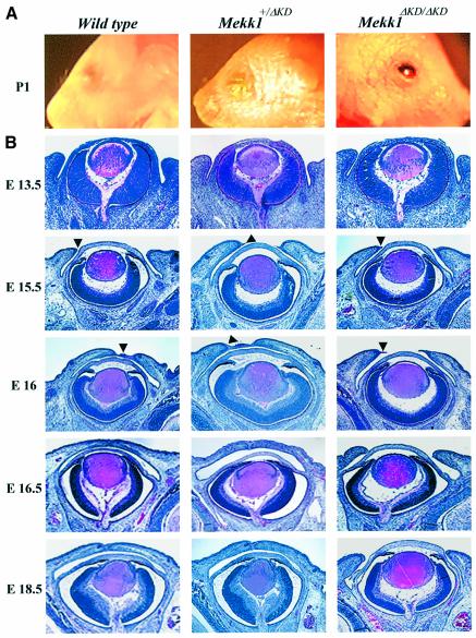
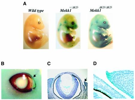
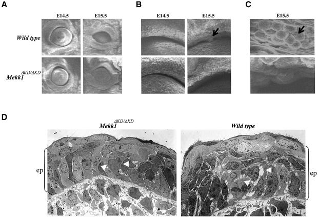
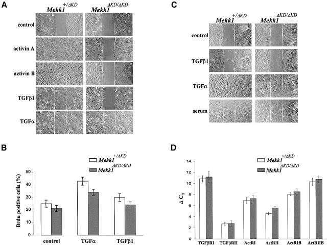
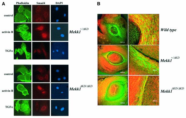
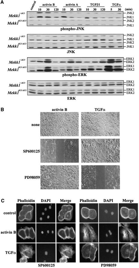
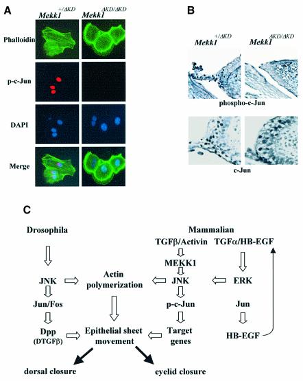
MEKK1-deficient mice show an eye open at birth phenotype caused by impairment in embryonic eyelid closure. MEK kinase 1 (MEKK1) is highly expressed in the growing tip of the eyelid epithelium, which displays loose cell-cell contacts and prominent F-actin fibers in wild-type mice, but compact cell contacts, lack of polymerized actin and a concomitant impairment in c-Jun N-terminal phosphorylation in MEKK1-deficient mice. In cultured keratinocytes, MEKK1 is essential for JNK activation by TGF-beta and activin, but not by TGF-alpha. MEKK1-driven JNK activation is required for actin stress fiber formation, c-Jun phosphorylation and cell migration. However, MEKK1 ablation does not impair other TGF-beta/activin functions, such as nuclear translocation of Smad4. These results establish a specific role for the MEKK1-JNK cascade in transmission of TGF-beta and activin signals that control epithelial cell movement, providing the mechanistic basis for the regulation of eyelid closure by MEKK1. This study also suggests that the signaling mechanisms that control eyelid closure in mammals and dorsal closure in Drosophila are evolutionarily conserved.


Figures

None
Fig. 1. MEKK1 ablation causes impairment in embryonic eyelid closure. (A) Photograph of the eyes of wt (left panel), Mekk1+/ΔKD (middle panel) and Mekk1ΔKD/ΔKD mice (right panel) at postnatal day 1, showing the EOB phenotype in Mekk1ΔKD/ΔKD mice. (B) H&E staining the coronal eye sections from wt (left panels), Mekk1+/ΔKD (middle panels) and Mekk1ΔKD/ΔKD (right panels) fetuses of various gestational ages (E13.5–E18.5). Arrowheads point to the developing eyelid tip and impaired eyelid closure is observed in Mekk1ΔKD/ΔKD fetuses at E16.
None
Fig. 2. MEKK1 expression during embryonic development. (Awt, Mekk1+/ΔKD and Mekk1ΔKD/ΔKD fetuses of E15.5 were stained for β-galactosidase expression. Photographs were taken at 7.5× magnification. (B) β-galactosidase expression in the eye of E15.5 Mekk1ΔKD/ΔKD fetuses. The arrow indicates the leading edge of the developing eyelid, photographed at 15× magnification. Coronal sections of X-gal-stained E15.5 fetuses, counterstained with hematoxylin and photographed at (C) 100× and (D) 200× magnification. β-galactosidase expressing cells (blue) were observed in a variety of eye tissues, including eyelid, conjunctiva, iris and ciliary body, retina and lens epithelium.
None
Fig. 3. MEKK1 ablation affects the eyelid epithelium morphology. Eye samples of wt and Mekk1ΔKD/ΔKD fetuses at the indicated ages were studied by scanning electron microscopy. Photographs taken at (A) low (60×), (B) medium (400×) and (C) high (1200×) magnification showing clumps of round cells along the eyelid margin (arrows) present in wt but not in Mekk1ΔKD/ΔKD E15.5 fetuses. (D) Transmission electron microscopy of the eyelid tip epithelium from wt and Mekk1ΔKD/ΔKD E15.5 fetuses. The wt eyelid epithelium (ep), delineated by the brackets, is thicker than its Mekk1ΔKD/ΔKD counterpart. The epithelium of the wt eyelid displays reduced cell–cell contacts and increased intercellular spaces (white arrowheads). In contrast, the epithelium of Mekk1ΔKD/ΔKD eyelids displays tight epithelial cell interactions (white arrowheads).
None
Fig. 4. MEKK1 is required for keratinocyte migration induced by TGF-β/activin. Confluent monolayers of mouse epidermal keratinocytes (A) or dermal fibroblasts (C) were subjected to in vitro wound-healing assays in medium without growth factors (control) or with activin A (5 ng/ml), activin B (5 ng/ml), TGF–β1 (10 ng/ml), TGF-α (10 ng/ml) or fetal calf serum (5%), as indicated. Photographs were taken immediately and 24 h after wounding; only the 24 h time point is shown. (BMekk1+/ΔKD and Mekk1ΔKD/ΔKD keratinocytes were labeled with BrdU and BrdU-positive cells were identified by indirect immunofluorescence. A total of 200 cells per experimental condition were examined and error bars indicate 95% confidence limits. (D) Total RNA isolated from Mekk1+/ΔKD and Mekk1ΔKD/ΔKD keratinocytes were examined by real-time RT–PCR for the expression of TGF-β/activin receptors, as indicated. Each experiment was performed in triplicate and the ordinate (ΔCT) represents the number of cycles needed to reach an arbitrary amplification threshold value normalized to GAPDH mRNA in the same sample.
None
Fig. 5. MEKK1 controls actin stress fiber formation in epidermal keratinocytes and in the developing eyelid epithelium. (AMekk1+/ΔKD and Mekk1ΔKD/ΔKD keratinocytes were maintained in growth-factor-free medium for 24 h before treatment with activin B (5 ng/ml) and TGF-α (10 ng/ml) for 2 h. Immunostaining was performed with FITC–phalloidin (green) for F-actin, anti-Smad4 and Alexa Fluor (red) for Smad4 and DAPI (blue) for nuclei. Activin B-induced actin stress fiber formation takes place in Mekk1+/ΔKD, but not in Mekk1ΔKD/ΔKD keratinocytes whereas, Smad4 nuclear translocation is unaffected by MEKK1 ablation. Photographs were taken at 600× magnification under fluorescence light. (B) Whole-mount staining of the eyes from wt, Mekk1+/ΔKD and Mekk1ΔKD/ΔKD fetuses at E15.5 was performed using FITC–phalloidin and propidium iodide for F–actin and DNA, respectively. The FITC–phalloidin binding to F-actin is clearly decreased in eyelids of Mekk1ΔKD/ΔKD fetuses. Images were captured by laser scanning microscope at low (left panels) and higher (right panels) magnifications.
None
Fig. 6. The TGF-β/activin-induced MEKK1–JNK pathway is required for actin polymerization and epithelial cell migration. (A) JNK activation by TGF-β/activin is mediated through MEKK1. The Mekk1+/ΔKD and Mekk1ΔKD/ΔKD keratinocytes were treated by various growth factors for the indicated times and cell lysates were assessed for phosphorylation and expression of JNK and ERK by western blotting using specific antibodies. JNK phosphorylation induced by TGF-β/activin was completely abolished by MEKK1 ablation, while ERK phosphorylation induced by TGF-α and TGF-β/activin was only marginally affected. Epithelial cell migration (B) and actin stress fiber formation (C) induced by activin B require JNK, while the response to TGF-α requires ERK. Mekk1+/ΔKD keratinocytes were deprived of growth factors for 24 h and pretreated with the JNK inhibitor SP600125 (5 µM) and the MEK inhibitor PD98059 (5 µM) for 0.5 h. The cells were cultured in medium without growth factors (control) or with either TGF-α (10 ng/ml) or activin B (5 ng/ml) for (B) 24 h for the in vitro wound healing assay and (C) 2 h for detection of F-actin formation by fluorescence staining. Wound closure and actin stress fiber formation induced by activin B were blocked by the JNK inhibitor, while the response to TGF-α was prevented by the ERK inhibitor.
None
Fig. 7. Involvement of c-Jun N-terminal phosphorylation in activin signaling and eyelid development. (A) MEKK1 is required for activin-B-induced c-Jun N-terminal phosphorylation in keratinocyte. Mekk1+/ΔKD and Mekk1ΔKD/ΔKD keratinocytes were maintained in growth-factor-free medium for 24 h, followed by treatment with activin B (5 ng/ml) for 2 h. Immunostaining was carried out using anti-phospho-c-Jun (S63), FITC–phalloidin and DAPI for c-Jun phosphorylation, F-actin and nuclei, respectively. Activin-B-induced c-Jun phosphorylation and F-actin are both abolished by MEKK1 ablation. (B) Eye sections of E15.5 Mekk1+/ΔKD and Mekk1ΔKD/ΔKD fetuses were immunostained with anti-phospho-c-Jun (S63) (top panels) or anti-c-Jun (bottom panels). Many cells in the eyelid epithelium of Mekk1+/ΔKD fetuses show positive nuclear phospho-c-Jun staining, while only a few phospho-c-Jun-positive cells are detected in Mekk1ΔKD/ΔKD eyelids. c-Jun-positive cells are detected at the similar frequencies in the eyelid epithelia of Mekk1+/ΔKD and Mekk1ΔKD/ΔKD. (C) An evolutionarily conserved JNK pathway regulates mammalian eyelid closure and Drosophila dorsal closure. In mammalian eyelid closure, one pathway involves the MEKK1–JNK cascade, required for receiving and transmitting the TGF-β/activin signal in the control of actin polymerization and c-Jun phosphorylation which is important for epithelial sheet movement. Another pathway is c-Jun controlled expression of HB-EGF, which in turn may activate the EGFR–ERK pathway that also leads to actin polymerization and epithelial sheet movement. In Drosophila, a parallel regulatory mechanism, involving JNK, c-Jun and TGF-β, controls dorsal epithelium actin polymerization and closure. The MEKK1–JNK and the EGFR–ERK pathways are likely mammalian equivalents of the Drosophila JNK-AP-1-TGF-β pathway, representing an evolutionarily conserved signaling mechanism that controls epithelial sheet movement and tissue closure across species.

Similar articles

Cited by

References

    1. Ashrafi S.H., Atassi,B., Erickson,R. and Sabet,T. (1993) Migration of epithelium during phenytoin-dependent gingival overgrowth in mice. Scanning Microsc., 7, 1247–1253. - PubMed
    1. Bakin A.V., Tomlinson,A.K., Bhowmick,N.A., Moses,H.L. and Arteaga,C.L. (2000) Phosphatidylinositol 3-kinase function is required for transforming growth factor beta-mediated epithelial to mesenchymal transition and cell migration. J. Biol. Chem., 275, 36803–36810. - PubMed
    1. Beck P.L., Rosenberg,I.M., Xavier,R.J., Koh,T., Wong,J.F. and Podolsky,D.K. (2003) transforming growth factor-beta mediates intestinal healing and susceptibility to injury in vitro and in vivo through epithelial cells. Am. J. Pathol., 162, 597–608. - PMC - PubMed
    1. Berkowitz E.A. et al. (1996) Characterization of the mouse transforming growth factor alpha gene: its expression during eyelid development and in waved 1 tissues. Cell Growth Differ., 7, 1271–1282. - PubMed
    1. Birk D.E. and Mayne,R. (1997) Localization of collagen types I, III and V during tendon development. Changes in collagen types I and III are correlated with changes in fibril diameter. Eur. J. Cell Biol., 72, 352–361. - PubMed

Publication types

MeSH terms