. 2003 Sep 1;22(17):4478-88.
doi: 10.1093/emboj/cdg434.

Progression through key stages of haemopoiesis is dependent on distinct threshold levels of c-Myb

Affiliations

Progression through key stages of haemopoiesis is dependent on distinct threshold levels of c-Myb

Nikla Emambokus et al. EMBO J. .

Abstract

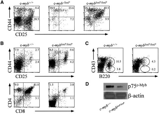
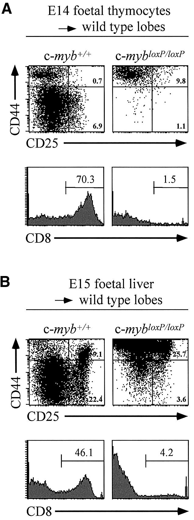
The c-Myb transcription factor is expressed in immature haemopoietic cells and at key stages during differentiation. Loss of the c-myb gene results in embryonic lethality because mature blood cells fail to develop, although commitment to definitive haemopoiesis occurs. We have generated a knockdown allele of c-myb, expressing low levels of the protein, which has enabled us to investigate further the involvement of c-Myb in haemopoiesis. Low levels of c-Myb are sufficient to allow progenitor expansion but, importantly, we show that progression of progenitors towards terminal differentiation is significantly altered. Suboptimal levels of c-Myb favour differentiation of macrophage and megakaryocytes, while higher levels seem to be particularly important in the control of erythropoiesis and lymphopoiesis. We provide evidence that the transition from the CFU-E to erythroblasts is critically dependent on c-Myb levels. During thymopoiesis, c-Myb appears to regulate immature cell numbers and differentiation prior to expression of CD4 and CD8. Overall, our results point to a complex involvement of c-Myb in the regulation of proliferation and commitment within the haemopoietic hierarchy.


Figures

None
Fig. 1. Generation of a conditional c-myb allele. The targetting vector is shown together with the organization of the wild-type c-myb gene. A neomycin resistance cassette (neo) for positive selection and the HSV-1 thymidine kinase gene (tk) for negative selection were introduced into intron 6 and just downstream of exon 9 respectively. The neo cassette was flanked by Flp recognition sites (open arrowheads). LoxP sites (filled arrowheads) were introduced into introns 2 and 6. The vertical black boxes represent exons. Relevant restriction endonuclease sites are indicated (E, EcoRI; H, HindIII and Sp, SpeI). The fragments used to probe Southern blots are indicated (external, neo and deletion probes).
None
Fig. 2. Insertion of the neoR cassette into exon 6 of the c-myb gene results in a knockdown allele that affects development. (A) Wild-type, c-myb–/– and c-myb–/loxP E15 embryos and dissected fetal livers. (B) Wild-type, c-mybloxP/loxP and c-myb–/loxP E15 embryos and dissected fetal livers from E15 and E14 embryos. (C) Wild-type and c-mybloxP/loxP littermates 6 days after birth. (D) c-mybloxP/loxP mice have a reduced lifespan. The graph represents the survival of 23 wild-type compared with 23 c-mybloxP/loxP mice derived from 15 litters. (E) The c-mybloxP allele produces low levels of full-length c-Myb protein. Western blot analysis of protein extracts of fetal livers from E11 embryos probed with a c-Myb specific monoclonal antibody. Ponceau staining of the filter immediately after transfer indicated that all tracks contained equivalent amounts of protein.
None
Fig. 3. Low levels of c-Myb permit haemopoietic progenitor development but affect commitment and differentiation. (A) The c-myb genotype affects fetal liver cellularity. Touch preparations of E15 fetal livers were stained with May–Grünwald–Giemsa. (B) Low level c-Myb expression leads to a relative accumulation of progenitor cells. Flow cytometric analysis of fetal liver cells from wild-type, c-mybloxP/loxP, c-myb–/loxP and c-myb–/– E14 embryos stained with anti-c-Kit–PE (upper panels) or anti-CD34–biotin followed by streptavidin–PE (lower panels). The bar indicates the percentage of positive cells relative to staining obtained using an isotype control. (C and D) Progenitors are functional in the presence of lower than normal levels of c-Myb. Fetal liver cells (4 × 10⁴) from E13 or E15 embryos were plated in duplicate in methycellulose to allow growth and identification of all myeloid types. Each determination was repeated at least three times. The data is presented as the total colony number (C) for E13 or E15 derived fetal liver and as the number of individual colony types obtained from E13 cells (D). (E) Mixed colonies differentiate more rapidly from c-myb–/loxP fetal liver progenitors. Cytospins of day 4 colonies from a CFU assay of E13 fetal livers were stained with May-Grünwald–Giemsa. Typical examples of macrophage (m), megakaryocytes (mk) and neutrophils (n) as well as more immature cells (i) are indicated. (F) Megakaryopoiesis is increased in vivo. Flow cytometric analysis of fetal liver cells from wild-type and c-myb–/loxP E13 embryos stained with anti-CD41 followed by streptavidin–PE and anti-IgG1–FITC.
None
Fig. 4. Erythroid differentiation is perturbed in the presence of reduced levels of c-Myb. (A and B) Flow cytometric analysis of fetal liver cells from wild-type, c-mybloxP/loxP, c-myb–/loxP and c-myb–/– embryos stained with Ter119–biotin and anti-CD71 followed by streptavidin–PE and anti-IgG1–FITC. The profiles in (A) are derived from E15 embryos. The summary histogram in (B) includes data from E15 embryos and also similar stainings of fetal livers from wild-type and c-mybloxP/loxP E14 embryos. (C and D) Culture of wild-type and c-mybloxP/loxP E14 fetal liver cells under conditions favouring expansion of erythroid precursors, maintaining a cell concentration of 1–3 × 10⁶/ml. (C) The cumulative cell number. (D) Cultured cells were stained with Ter119-biotin and anti-CD71 as described in (A) at day 8.
None
Fig. 5. Lymphoid differentiation is particularly sensitive to the level of c-Myb. (A) Flow cytometric analysis of fetal thymus cells from wild-type, c-mybloxP/loxP and c-myb–/loxP E15 embryos stained with anti-CD44–PE and anti-CD25–FITC. (B) Flow cytometric analysis of thymus cells from wild-type and c-mybloxP/loxP neonates stained with anti-CD44–PE and anti-CD25–FITC (upper panels) and anti-CD4–PE and anti-CD8–FITC (lower panels). (C) Flow cytometric analysis of bone marrow cells from wild-type and c-mybloxP/loxP neonates stained with anti-CD43–PE and anti-B220–FITC. (D) Western blot analysis of protein extracts of sorted CD4+8+ 3 month-old thymocytes probed with monoclonal antibodies specific for c-Myb and β-actin as a control for loading.
None
Fig. 6. Development of c-myb knockdown thymocytes in organ culture. Flow cytometric analysis of CD44 and CD25 expression (upper panels) and CD8 expression (lower panels) on cells derived from deoxyguanosine-treated thymic lobes seeded and subsequently cultured with thymocyte progenitors. (A) Cells from wild-type or c-mybloxP/loxP E14 thymuses were cultured with thymic lobes for 7 days. (B) Fragments of fetal liver from wild-type or c-mybloxP/loxP E15 embryos were incubated with thymic lobes separated by a 0.3 µm filter for 24 h. The fetal liver was then removed and the lobes cultured for an additional 16 days.

Similar articles

Cited by

References

    1. Allen R.D., Bender,T.P. and Siu,G. (1999) c-Myb is essential for early T cell development. Genes Dev., 13, 1073–1078. - PMC - PubMed
    1. Allen R.D., Kim,H.K., Sarafova,S.D. and Siu,G. (2001) Negative regulation of CD4 gene expression by a HES-1-c-Myb complex. Mol. Cell. Biol., 21, 3071–3082. - PMC - PubMed
    1. Clarke M.F., Kukowska-Latallo,J.F., Westin,E., Smith,M. and Prochownik,E.W. (1988) Constitutive expression of a c-myb cDNA blocks friend murine erythroleukemia cell differentiation. Mol. Cell. Biol., 8, 884–892. - PMC - PubMed
    1. Clarke D., Vegiopoulos,A., Crawford,A., Mucenski,M., Bonifer,C. and Frampton,J. (2000) In vitro differentiation of c-myb–/– ES cells reveals that the colony forming capacity of unilineage macrophage precursors and myeloid progenitor commitment are c-Myb independent. Oncogene, 19, 3343–3351. - PubMed
    1. DeKoter R.P. and Singh,H. (2000) Regulation of B lymphocyte and macrophage development by graded expression of PU.1. Science, 288, 1439–1441. - PubMed

Publication types

MeSH terms