. 2003 Sep 1;22(17):4555-65.
doi: 10.1093/emboj/cdg433.

Group II intron mobility using nascent strands at DNA replication forks to prime reverse transcription

Affiliations

Group II intron mobility using nascent strands at DNA replication forks to prime reverse transcription

Jin Zhong et al. EMBO J. .

Abstract

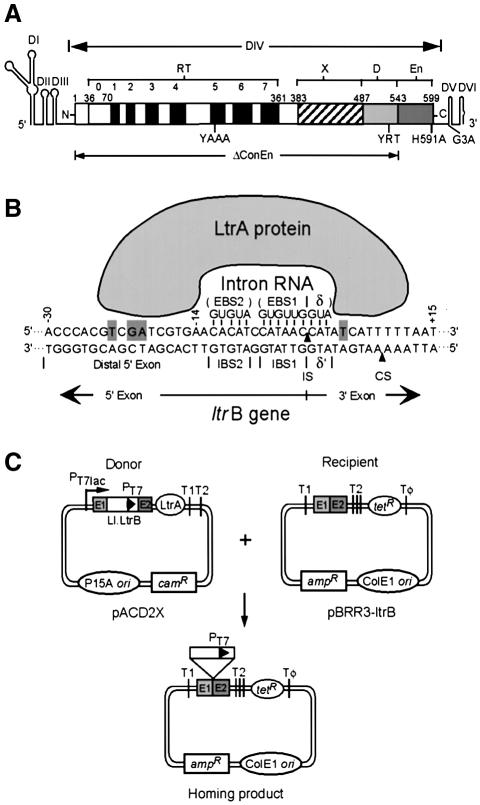
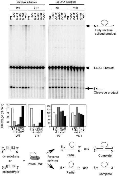
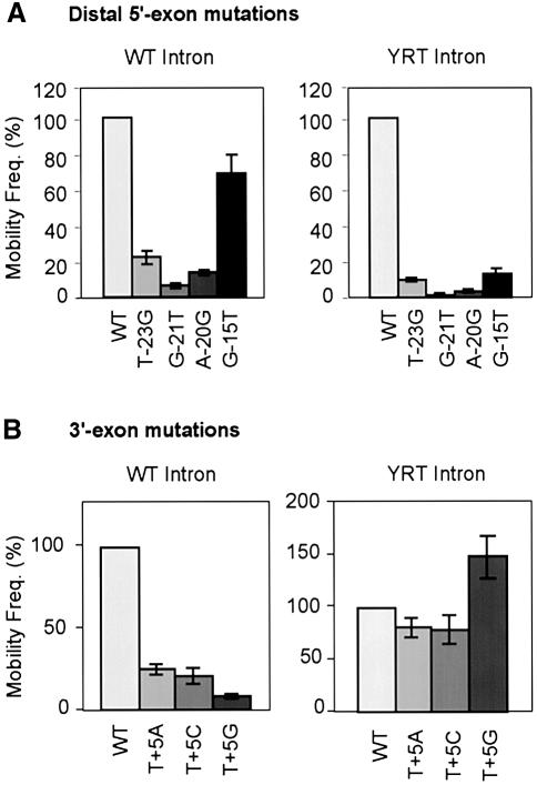
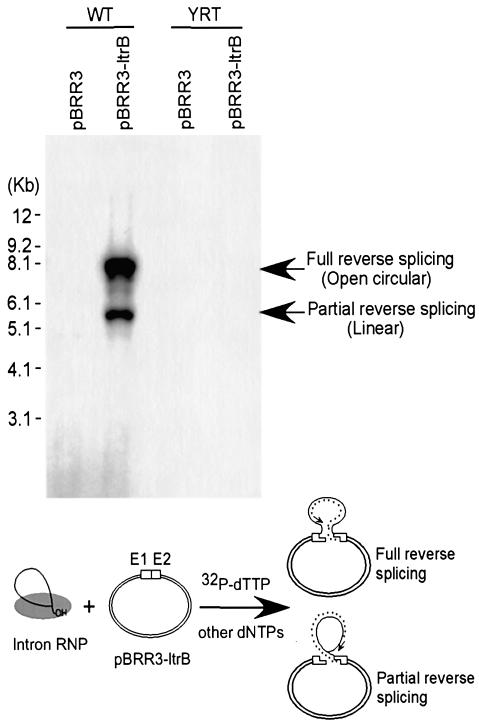
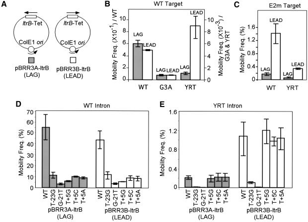
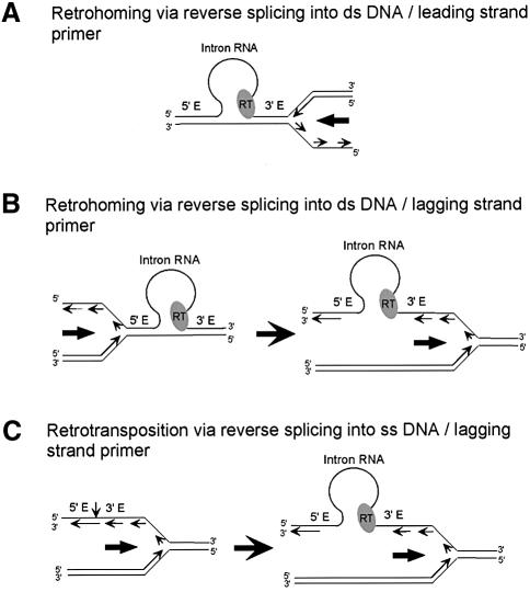
The Lactococcus lactis Ll.LtrB group II intron uses a major retrohoming mechanism in which the excised intron RNA reverse splices into one strand of a DNA target site, while the intron-encoded protein uses a C-terminal DNA endonuclease domain to cleave the opposite strand and then uses the cleaved 3' end as a primer for reverse transcription of the inserted intron RNA. Here, experiments with mutant introns and target sites indicate that Ll.LtrB can retrohome without second-strand cleavage by using a nascent strand at a DNA replication fork as the primer for reverse transcription. This mechanism connecting intron mobility to target DNA replication may be used by group II intron species that encode proteins lacking the C-terminal DNA endonuclease domain and for group II intron retrotransposition to ectopic sites.


Figures

None
Fig. 1. The Ll.LtrB group II intron, DNA target site interactions, and plasmid-based intron mobility assay. (A) Ll.LtrB intron. The intron is shown as a thin line, with RNA secondary structure domains DI–VI drawn schematically. The IEP, denoted LtrA protein, is shown as an open box. The protein contains an N-terminal RT domain, with conserved RT sequence motifs 0–7; domain X associated with maturase activity; and C-terminal DNA-binding (D) and DNA endonuclease (En) domains. The locations of mutations YAAA, YRT, H591A, G3A and ΔConEn are shown below. (B) Mechanism of DNA target site recognition. The DNA target site in the ltrB gene is recognized by an RNP complex containing the IEP (LtrA protein) and excised intron lariat RNA, with both protein interactions and base pairing of the intron RNA contributing to DNA target site recognition. Bases identified as being recognized directly by the LtrA protein (Singh and Lambowitz, 2001) are highlighted with gray backgrounds. The arrowheads pointing to the top and bottom strands indicate the intron-insertion site (IS) and second-strand cleavage site (CS), respectively. (C) Mobility assay. The intron-donor plasmid pACD2X is a CamR pACYC184-derivative (P15 replicon), which contains a 940-nt Ll.LtrB-ΔORF intron and flanking exons, with a phage T7 promoter inserted near the 3′ end of the intron. The intron is expressed from a T7lac promoter, with the IEP (LtrA protein) expressed from a position just downstream of the 3′ exon. The recipient plasmid pBRR3-ltrB is an AmpR pBR322-derivative, which contains the Ll.LtrB target site (ligated E1-E2 sequence of the ltrB gene) cloned upstream of a promoterless tetR gene. The insertion of the intron into the target site activates the expression of the tetR gene, yielding TetR+AmpR colonies. T1, T2 and Tφ are E. coli rrnB T1, T2 and phage T7 transcription terminators, respectively.
None
Fig. 2. Reverse splicing assay with double- and single-stranded DNA target sites. Reverse splicing was assayed by incubating wild-type or YRT mutant RNP particles with 5′-top-strand labeled double- or single-stranded 70mer DNAs containing the wild-type target sequence (positions –35 to +35) or the indicated mutations. The products were analyzed in a denaturing 6% polyacrylamide gel, which was dried and quantitated with a PhosphorImager. Labeled products are indicated to the right of the gel, with the position of the label indicated by an asterisk. The schematic at the bottom diagrams reverse splicing of the intron RNA into double- or single-stranded DNA. Partial reverse splicing results in cleaved 5′-exon (the predominant product) and intron lariat linked to 3′-exon (not labeled), while complete reverse splicing results in insertion of linear intron RNA between the two DNA exons. The bar graphs show reverse splicing activity relative to the wild-type DNA target site based on quantitation of the partially and fully reverse spliced products.
None
Fig. 3. Mobility assays with wild-type and mutant DNA target sites. Mobility assays were carried out as described in Figure 1 and Materials and methods, with donor plasmids expressing the wild-type (WT) or YRT mutant introns and recipient plasmids containing wild-type or indicated mutant Ll.LtrB target sites (positions –30 to +15). (A) Distal 5′-exon mutations T-23G, G-21T, A-20G and G-15T; (B) 3′-exon mutations T+5A, T+5C and T+5G. The bar charts show mobility frequencies for mutant DNA target sites relative to the wild-type DNA target site assayed in parallel. Values are the mean ± SD of three independent experiments.
None
Fig. 4. Target DNA-primed reverse transcription assay. Wild-type and YRT mutant RNP particles were incubated with recipient plasmid pBRR3-ltrB, which contains the Ll.LtrB target site (positions –30 to +15), or control plasmid pBRR3, which lacks the Ll.LtrB target site, in the presence of [α-32P]dTTP and other dNTPs. Products were analyzed in a 0.7% agarose gel, which was dried and scanned with a PhosphorImager. DNA marker positions are shown to the left, and the products are identified to the right based on previous characterization (Saldanha et al., 1999). The schematic at the bottom shows the TPRT reaction, with products resulting from cDNA synthesis after partial or complete reverse splicing of the intron RNA into the DNA target site. The small asterisks indicate cDNA with the arrow indicating the direction of reverse transcription.
None
Fig. 5. Mobility assays with DNA target sites cloned in opposite orientations relative to the direction of plasmid replication. (A) Recipient plasmids pBRR3A-ltrB and pBRR3B-ltrB. The plasmids contain the ltrB target site/tetR cassette cloned in opposite orientations denoted LAG or LEAD, depending on whether the nascent lagging or leading DNA strands could be used as a primer for reverse transcription of the inserted intron RNA. (B) Mobility assays with the wild-type (WT), G3A or YRT mutant introns and recipient plasmids pBRR3A-ltrB and pBRR3B-ltrB. The scale on the left is for the wild-type intron, and the scale on the right is for the G3A and YRT mutant introns. (C) Mobility assays with the wild-type Ll.LtrB intron and recipient plasmids containing different orientations of mutant DNA target site E2m, which has 3′-exon mutations that inhibit second-strand cleavage but not reverse splicing (see Materials and methods). (D and E) Mobility assays with the wild-type (D) or YRT mutant (E) introns and recipient plasmids containing different orientations of mutant DNA target sites having distal 5′-exon (T-23G and G-21T) or 3′-exon (T+5G, T+5C and T+5A) mutations. Mobility assays were carried out as described in Figure 1 and Materials and methods. Values are the mean ± SD of three independent experiments.
None
Fig. 6. Models for group II intron mobility mechanisms using nascent leading and lagging-strand DNA primers at a replication fork. (A and B) Retrohoming via reverse splicing into double-stranded DNA, followed by use of a nascent leading strand (A) or lagging strand (B) as a primer for reverse transcription of the inserted intron RNA. After reverse splicing into the leading-strand template, the intron RNP is positioned to directly use the nascent leading strand as a primer prior to passage of the replication fork. In contrast, after reverse splicing into the lagging-strand template, the DNA replication machinery must traverse the inserted intron RNP prior to use of a lagging-strand primer. The observed leading-strand preference may reflect interference between the replication machinery and inserted intron RNP during passage of the replication fork in the lagging-strand orientation. (C) Retrotransposition of the Ll.LtrB intron in L.lactis. The group II intron RNP reverse splices into single-stranded DNA behind the replication fork and uses a nascent lagging strand to prime reverse transcription (see also Ichiyanagi et al., 2002). The models account for the opposite strand preferences of group II intron retrohoming and retrotransposition and for the finding that most group II intron retrotransposition sites lack distal 5′-exon sequences shown here to be required for efficient reverse splicing into double-stranded DNA. Some retrotransposition events also occur via reverse splicing into the leading-strand template and may use a nascent leading strand or non-specifically nicked DNA strand as primer.

Similar articles

Cited by

References

    1. Aizawa Y., Xiang,Q., Lambowitz,A.M. and Pyle,A.M. (2003) The pathway for DNA recognition and RNA integration by a group II intron retrotransposon. Mol. Cell, 11, 795–805. - PubMed
    1. Bazaral M. and Helinski,D.R. (1970) Replication of a bacterial plasmid and an episome in Escherichia coli. Biochemistry, 9, 399–406. - PubMed
    1. Belfort M., Derbyshire,V., Parker,M.M., Cousineau,B. and Lambowitz,A.M. (2002) Mobile introns: pathways and proteins. In Craig,N.L., Craigie,R., Gellert,M. and Lambowitz,A.M. (eds), Mobile DNA II. ASM Press Publishers, Washington, DC, pp. 761–783.
    1. Dai L. and Zimmerly,S. (2002) Compilation and analysis of group II intron insertions in bacterial genomes: evidence for retroelement behavior. Nucleic Acids Res., 30, 1091–1102. - PMC - PubMed
    1. Dickson L., Huang,H.R., Liu,L., Matsuura,M., Lambowitz,A.M. and Perlman,P.S. (2001) Retrotransposition of a yeast group II intron occurs by reverse splicing directly into ectopic DNA sites. Proc. Natl Acad. Sci. USA, 98, 13207–13212. - PMC - PubMed

Publication types