. 2003 Sep 1;22(17):4566-76.
doi: 10.1093/emboj/cdg429.

Full-length archaeal Rad51 structure and mutants: mechanisms for RAD51 assembly and control by BRCA2

Affiliations

Full-length archaeal Rad51 structure and mutants: mechanisms for RAD51 assembly and control by BRCA2

David S Shin et al. EMBO J. .

Abstract

To clarify RAD51 interactions controlling homologous recombination, we report here the crystal structure of the full-length RAD51 homolog from Pyrococcus furiosus. The structure reveals how RAD51 proteins assemble into inactive heptameric rings and active DNA-bound filaments matching three-dimensional electron microscopy reconstructions. A polymerization motif (RAD51-PM) tethers individual subunits together to form assemblies. Subunit interactions support an allosteric 'switch' promoting ATPase activity and DNA binding roles for the N-terminal domain helix-hairpin-helix (HhH) motif. Structural and mutational results characterize RAD51 interactions with the breast cancer susceptibility protein BRCA2 in higher eukaryotes. A designed P.furiosus RAD51 mutant binds BRC repeats and forms BRCA2-dependent nuclear foci in human cells in response to gamma-irradiation-induced DNA damage, similar to human RAD51. These results show that BRCA2 repeats mimic the RAD51-PM and imply analogous RAD51 interactions with RAD52 and RAD54. Both BRCA2 and RAD54 may act as antagonists and chaperones for RAD51 filament assembly by coupling RAD51 interface exchanges with DNA binding. Together, these structural and mutational results support an interface exchange hypothesis for coordinated protein interactions in homologous recombination.


Figures

None
Fig. 1. Sequence, secondary structure, conservation, fold, residue function and domain architecture for the RAD51 protein family. (A) Alignment of RAD51 homologs from P.furiosus (PfRad51), H.sapiens (HsRAD51) and S.cerevisiae (ScRAD51). P and H under the sequence refer to PfRad51 and HsRAD51 key residues or mutations used in this study, while 2, 4 and 5 refer to ScRAD51 mutations that influence binding to ScRAD52, ScRAD54 or ScRAD55, respectively. B refers to HsRAD51 residues that bind BRC4. Triangles indicate contact residues between one subunit (blue) and its adjacent neighbor (black), or between heptamers (orange). (B) The N-terminal domain (ND, top) and ATPase domain (AD, bottom) of PfRad51 are connected by an elbow linker. Key motifs are colored according to the labels in (A). (C) Topology of a PfRad51 subunit shows conservation of the RecA-AD fold (Story et al., 1992). (D) Overlay of PfRad51 (orange) with HsRAD51-ND (purple) and HsRAD51-AD (green) reveals strong structural conservation. (E) Overlay of PfRad51-AD (orange) and EcRecA-AD (green) reveals a conserved ATPase fold with additional PfRad51-ND (red) and EcRec-AC (dark green) DNA binding domains positioned at opposite poles. (F) Organization of the recombinase family protein sequences. Regions of homology among RAD51-NDs, and RAD51-ADs and RecA-AD are colored red and yellow, respectively. Walker A and B motifs are green. Non-homologous regions are white or blue.
None
Fig. 2. PfRad51 polymeric assembly and polymerization motif. (A) Electron micrographs of PfRad51 reveal heptameric ring structures (scale bar, 20 nm). (B) SAXS intensities from PfRad51 (circles) plotted against the momentum transfer Q and calculated profiles for heptameric (dotted line) and biheptameric (thin curve) models indicate a biheptameric ring assembly. Rigid-body refinement of two heptamers improved the fit into the experimental data (thick curve). (C) Electron pair distribution functions P(r) for the SAXS data (circles) and for the heptamer (dotted line), biheptamer (thin curve) and rigid-body refined biheptamer (thick curve) models support biheptameric assembly. (D) Interface between two adjacent ATPase domains oriented similarly to the boxed region in (E) showing the β03 inter-subunit β-sheet. The N-terminal domains have been removed for clarity. (E and F) Single PfRad51 heptamer (E, top view) and biheptamer (F, side view) models show 7-fold symmetric assembly. Sulfates (balls) denote the ATPase active site. (G) A polymerization motif is formed by β0 (98-MRA-100) of the inter-subunit β-sheet and buried Phe97 and Ala100 side chains. The adjacent subunits are yellow and white and in an orientation similar to that in (D). Composite omit 2FoFc density is contoured at 2σ (purple) and 4σ (pink) and hydrogen bonds are shown as dashed lines.
None
Fig. 3. The ATPase active site and mutational decoupling of ATPase and strand exchange activities. (A and B) Comparisons of the ATP active sites of the PfRad51 ring (yellow) and EcRecA helical filament (green) suggest a mechanism for conformation-induced allostery for ATP binding. Key side chains and ADP (purple) from the RecA structure are shown as balls and sticks. In the PfRad51 ring, hydrophobic interactions between α5 and α9 of adjacent subunits pull Arg181 (α9) up and away from the nucleotide base (A). This arrangement may also allow PfRad51 Ile342 (β8) to sterically hinder nucleotide binding (B). In helical EcRecA, the absence of Pro101 hydrophobic contacts with the adjacent subunit allows Tyr103 to stack with the nucleotide base (A). (C) Analysis of ATPase activity of wild-type PfRad51 and L1 region mutants, R251A and R251E, with no DNA, ssDNA or dsDNA reveals that all proteins hydrolyze ATP in a DNA-dependent manner. (D) The ability to form joint molecules between circular ssDNA and linear dsDNA is greatly diminished for the PfRad51 R251A and R251E mutants compared with wild-type protein, despite their ability to hydrolyze ATP. (E) The more robust activity of AfRad51 wild-type protein confirms the results in (D) by comparison with the activity of an analogous AfRad51 R228A mutation.
None
Fig. 4. PfRad51 and EcRecA helical filament models and the implied Rad51 HhH DNA binding site. (A) PfRad51 filament assembly with PfRad51-ADs (alternating in orange and green) placed from superposition with EcRecA-ADs from X-ray crystallography (B) (Story et al., 1992). (B) The helical EcRecA structure with EcRecA-ADs alternating orange and green with C-terminal domains (yellow) and ADP molecules (balls). The PfRad51-NDs (A; yellow) within the groove have opposite polarity to the EcRecA-CDs. (C) Independent rigid body docking of the PfRad51 crystal structure into 3D EM reconstruction density of the S.solfataricus Rad51 homolog bound to DNA retained the basic features of the model presented in (A) that was based upon crystallographic polymers. (D) Positive charges [2.0 kT/e (blue) to –2.0 kT/e (red)] for the L1 region implicated in DNA interaction are contributed largely by Arg251 residues (center). Asymmetry reflects L1 region flexibility. (E) The PfRad51 helical filament model has positive electrostatic potential for DNA binding within the ssDNA binding interior and for the HhH motifs. (F) A dsDNA model fits into the large outer groove of the PfRad51 filament when guided by HhH-containing protein:DNA X-ray cocrystal structures. This suggests a method for RAD51 to bind ssDNA internally and dsDNA externally for homology search reactions, in which pairing may occur within channels.
None
Fig. 5. Molecular basis for BRCA2 regulation of RAD51 function. (A) PfRad51 with adjacent subunit interface polypeptide (green), showing the key subunit interface polymerization motif elements β0 and Phe97. (B) Superposition of HsRAD51-AD:BRC4 (HsRAD51-AD not shown) onto the PfRad51 structure (ribbons) reveals that the BRC4 repeat (green) occupies the same area as PfRad51-ND (not shown) and mimics β0 to disrupt the inter-subunit β-sheet between RAD51 subunits. (C) The PfRad51 van der Waals surface (blue) with the foremost subunit (coil) overlaid with BRC4 (orange) from HsRAD51-AD:BRC4 shows how RAD51-ND displacement and disassembly of the ring involves intercalation of the BRC repeat between RAD51 subunits. (D) Critical components of the BRC4 repeat (orange) for mimicry of the RAD51 polymerization motif. Phe1524 and Ala1527 match the adjacent PfRad51 subunit residues Phe97 and Ala100 (blue). (E) Binding of HsRAD51, PfRad51 and the PfRad51 E219S/D220A/D267M mutant to BRCA2 BRC3/BRC4 repeats establishes the additional critical components for RAD51:BRC repeat binding. In each triplet, we show the amount of input protein (first lane), a negative control of GST alone (second lane) and binding to a GST-BRC3/4 fusion protein (third lane). (F and G) BRC-dependent disassembly and targeting of mutant PfRad51 shown by fluorescence. A GFP-PfRad51 E219S/D220A/D267M mutant is targeted to dsDNA breaks in human 293T cells forming nuclear foci following γ-irradiation (F). The ability to form foci is abolished in the presence of BRC repeats 3 and 4 (G).
None
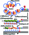
Fig. 6. Proposed model for BRCA2 coordination of RAD51 activities in HRR. (1) BRCA2 binds to RAD51 subunits within the ring (AD, brown; ND, red; elbow linker/β0, yellow arrow; β3, brown arrow) via BRC repeat mimicry of the RAD51 polymerization motif (β0 mimic, blue arrow). (2) BRC repeats disassemble the ring. (3) The RAD51:BRCA2 complex is recruited to a DSB. (4) BRCA2 helps displace RPA and binds the primary ssDNA substrate by its OB folds (5), and loads RAD51 onto DNA. The handoff reactions might be facilitated by attraction of DNA by the positively charged BRC repeat helical arches. The BRCA2 HTH domain (red) may bind dsDNA in cis at the ssDNA/dsDNA intra-DNA junction (3) or in trans to the dsDNA that later serves as the homologous DNA template (6) and the positively charged arch may also help to attract the dsDNA template.

Similar articles

Cited by

References

    1. Aihara H., Ito,Y., Kurumizaka,H., Terada,T., Yokoyama,S. and Shibata,T. (1997) An interaction between a specified surface of the C-terminal domain of RecA protein and double-stranded DNA for homologous pairing. J. Mol. Biol., 274, 213–221. - PubMed
    1. Aihara H., Ito,Y., Kurumizaka,H., Yokoyama,S. and Shibata,T. (1999) The N-terminal domain of the human Rad51 protein binds DNA: structure and a DNA binding surface as revealed by NMR. J. Mol. Biol., 290, 495–504. - PubMed
    1. Ariyoshi M., Nishino,T., Iwasaki,H., Shinagawa,H. and Morikawa,K. (2000) Crystal structure of the Holliday junction DNA in complex with a single RuvA tetramer. Proc. Natl Acad. Sci. USA, 97, 8257–8262. - PMC - PubMed
    1. Benson F.E., Stasiak,A. and West,S.C. (1994) Purification and characterization of the human Rad51 protein, an analogue of E.coli RecA. EMBO J., 13, 5764–5771. - PMC - PubMed
    1. Datta S., Prabu,M.M., Vaze,M.B., Ganesh,N., Chandra,N.R., Muniyappa,K. and Vijayan,M. (2000) Crystal structures of Mycobacterium tuberculosis RecA and its complex with ADP-AlF(4): implications for decreased ATPase activity and molecular aggregation. Nucleic Acids Res., 28, 4964–4973. - PMC - PubMed

Publication types

MeSH terms

Associated data