. 2003 Sep;77(18):9809-16.
doi: 10.1128/jvi.77.18.9809-9816.2003.

The cap region of topoisomerase I binds to sites near both ends of simian virus 40 T antigen

Affiliations

The cap region of topoisomerase I binds to sites near both ends of simian virus 40 T antigen

Rupa Roy et al. J Virol. 2003 Sep.

Abstract

Two independent binding sites on simian virus 40 (SV40) T antigen for topoisomerase I (topo I) were identified. One was mapped to the N-terminal domain (residues 83 to 160) by a combination of enzyme-linked immunosorbent assays (ELISAs) and glutathione S-transferase (GST) pull-down assays performed with various T antigen deletion mutants. The second was mapped to the C-terminal domain (residues 602 to 708). The region in human topo I that binds to both sites in T antigen was identified by ELISAs, GST pull-down assays, and double-hexamer binding assays with topo I deletion mutants. This region corresponds to a distinct domain on topo I known as the cap region that maps from residues 175 to 433. By combining these data with information about the structure of T-antigen double hexamers associated with origin DNA, we propose that the cap region of topo I associates specifically with both ends of the double hexamer bound to the SV40 origin to initiate DNA replication.


Figures

FIG. 1.
FIG. 1.
(Top) Map of topoisomerase I deletion mutants used in this study. Various fragments of human topo I were expressed either in bacteria (t) or in baculovirus-infected insect cells (v). The topo I polypeptides correspond to known topo I domains (40-42).
FIG.2.
FIG.2.
(Bottom) ELISA between WT T antigen and various topo I fragments. A total of 100 ng of purified WT topo I, topo 70, or topo 58 was bound to ELISA plates, followed by incubation with various amounts of WT T antigen as shown. The plates were washed and incubated with PAb101, followed by the addition of horseradish peroxidase-labeled anti-mouse IgG and peroxidase substrate. After the reactions were stopped with sulfuric acid, the absorbance at 490 nm was measured by using a plate reader. Control reactions were also performed without topo I but in the presence of all other components. The absorbance values from these wells were subtracted from the corresponding ones obtained in the presence of WT or mutant topo I.
FIG. 3.
FIG. 3.
GST-binding assay between WT T antigen and various topo I fragments. A total of 10 μl of glutathione-Sepharose or -agarose beads containing 18 μg of GST-tagged topo 70 or the molar equivalent of topo 31, topo 17, topo 10, or GST alone was incubated with 500 ng of WT T antigen (A) or the molar equivalent of 246-708 (B) or 1-246 (C) in the presence of 1 mM ethidium bromide. The beads were then washed and resuspended in electrophoresis sample buffer. Samples were loaded onto 10% acrylamide gels. The proteins were transferred to nitrocellulose, and the T-antigen polypeptides were detected with PAb101 (A and B) or PAb419 (C) in a Western blot assay. A small amount of WT T antigen and 246-708 was also added directly to the gels to serve as markers (last lanes).
FIG. 4.
FIG. 4.
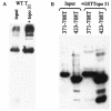
Origin DNA-binding assay with various topo I fragments. A total of 100 ng of GST-tagged topo 70 or the molar equivalent of various topo deletion mutants was incubated with 5 ng of circular origin DNA in the presence or absence of 400 ng of WT T antigen under replication conditions. After cross-linking, the samples were subjected to electrophoresis in a composite acrylamide agarose gel, and the protein complexes were transferred to nitrocellulose. topo I polypeptides were detected by reactivity with an anti-GST antibody in a Western blot assay. The positions of double hexamers (DH) are indicated and were determined by probing the same blots with an anti-T antibody (not shown).
FIG. 5.
FIG. 5.
(Top) Map of T antigen polypeptides used in this study. All of these proteins were expressed in baculovirus-infected insect cells. Nontagged polypeptides were purified by immunoaffinity chromatography, and GST-tagged proteins were purified by binding to glutathione-Sepharose beads.
FIG.6.
FIG.6.
(Bottom) ELISA between topo 70 and various N-terminal T antigen constructs. A total of 100 ng of topo 70 was first bound to microtiter plates. After a blocking step, various amounts of WT T antigen, as shown, or the molar equivalent of various N-terminal constructs of T antigen were added. After the wells were washed, PAb416 (which reacts with all T constructs used here) was added, followed by horseradish peroxidase-conjugated anti-mouse IgG. The absorbance at 490 nm was measured as described in Fig. 2. Controls without topo 70 were also performed for each T antigen construct, and these numbers were subtracted from the corresponding experimental numbers.
FIG. 7.
FIG. 7.
GST-binding assay between topo 31 and N-terminal T-antigen polypeptides and between small T antigen and topo I constructs. (A) A 10-μl portion of beads containing 2.5 μg of topo 31 was incubated with 500 ng of WT T antigen or the molar equivalent of various N-terminal T antigen constructs. The beads were washed and resuspended in electrophoresis sample buffer, and the samples were applied to a 13% acrylamide gel. Detection of the T-antigen polypeptides was performed with PAb419, which binds to the N-terminal region of T antigen. Samples of all polypeptides were loaded onto the gel to serve as markers of the input material (lanes on left). (B) Portions (10 μl) of beads containing 18 μg of GST-tagged topo 70 or the molar equivalent of topo 31, topo 17, topo 10, or GST alone were incubated with 150 ng of small T antigen (a gift of Kathy Rundell). Bound proteins were applied to a 13% acrylamide gel, and small T antigen on the membrane was detected with PAb419. A sample of small T antigen was loaded directly in the last lane.
FIG. 8.
FIG. 8.
GST-binding assay between topo 31 and C-terminal T antigen constructs. A total of 10 μl of agarose beads containing 2.5 μg of GST-topo 31 was incubated with 500 ng of WT T antigen (A) or the molar equivalent of either 371-708 or 423-708 T-antigen polypeptides (B). Bound T-antigen proteins were detected by reactivity with PAb101, which binds to the C-terminal end of T antigen. Samples of the input proteins are shown as indicated.
FIG. 9.
FIG. 9.
GST-binding assay between WT topo I and various C-terminal T antigen constructs. A total of 30 μl of Sepharose beads containing 8 μg of bound GST 110-708 or the molar equivalent of various C-terminal constructs or control GST beads was incubated with 1.8 μg of purified WT topo I. After the beads were washed, bound topo I was eluted with electrophoresis sample buffer and applied to an 8% gel. Detection was with 8G6 monoclonal antibody and horseradish peroxidase-conjugated anti-mouse IgG.
FIG. 10.
FIG. 10.
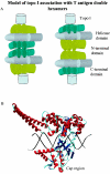
(A) Model of topo I associated with a T-antigen double hexamer. This model is based on the data presented by VanLoock et al. (48) and Valle et al. (47). Each hexamer of T antigen consists of three domains: an N-terminal DNA-binding domain, a central helicase domain, and a C-terminal domain. The hexamers are oriented either with their C-terminal domains facing each other (model on left) or with the N-terminal domains facing one another (model on right). topo I is shown binding to the N-terminal domains on left or to the C-terminal domains on right. (B) topo 31 is a distinct domain of topo I. This illustration is based on the topo 70 crystal structure by Rebindo et al. (32) and Stewart et al. (43) (pdb entry 1a36). The topo 31 domain (the cap region) is shown below the DNA.

Similar articles

Cited by

References

    1. Borowiec, J. A. 1992. Inhibition of structural changes in the simian virus 40 core origin of replication by mutation of essential origin sequences. J. Virol. 66:5248-5255. - PMC - PubMed
    1. Borowiec, J. A., F. B. Dean, P. A. Bullock, and J. Hurwitz. 1990. Binding and unwinding: how T antigen engages the SV40 origin of DNA replication. Cell 60:181-184. - PubMed
    1. Borowiec, J. A., F. B. Dean, and J. Hurwitz. 1991. Differential induction of structural changes in the simian virus 40 origin of replication by T antigen. J. Virol. 65:1228-1235. - PMC - PubMed
    1. Borowiec, J. A., and J. Hurwitz. 1988. Localized melting and structural changes in the SV40 origin of replication induced by T-antigen. EMBO J. 7:3149-3158. - PMC - PubMed
    1. Braun, K. A., Y. Lao, Z. He, C. J. Ingles, and M. S. Wold. 1997. Role of protein-protein interactions in the function of replication protein A (RPA): RPA modulates the activity of DNA polymerase alpha by multiple mechanisms. Biochemistry 36:8443-8454. - PubMed

Publication types

MeSH terms

Substances