. 2003 Sep;77(18):9969-78.
doi: 10.1128/jvi.77.18.9969-9978.2003.

Integrin-using rotaviruses bind alpha2beta1 integrin alpha2 I domain via VP4 DGE sequence and recognize alphaXbeta2 and alphaVbeta3 by using VP7 during cell entry

Affiliations

Integrin-using rotaviruses bind alpha2beta1 integrin alpha2 I domain via VP4 DGE sequence and recognize alphaXbeta2 and alphaVbeta3 by using VP7 during cell entry

Kate L Graham et al. J Virol. 2003 Sep.

Abstract

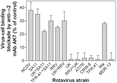
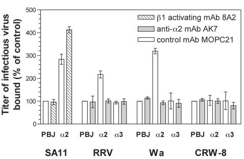
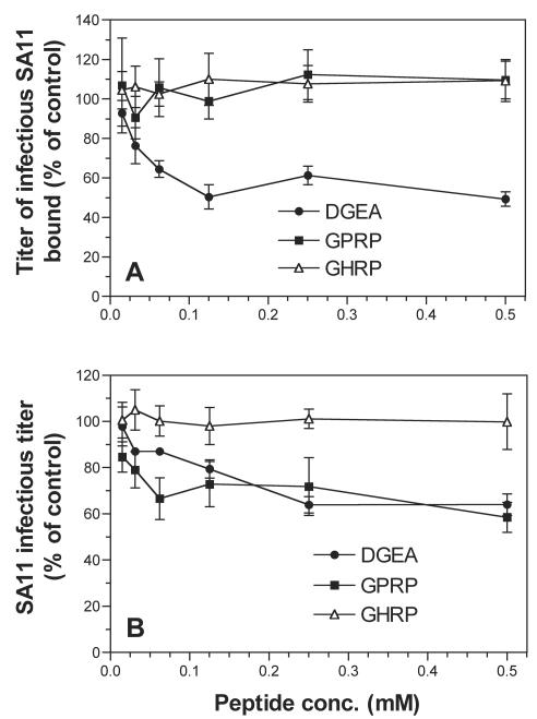
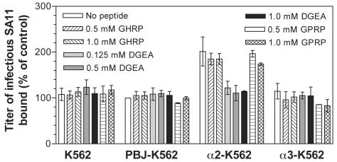
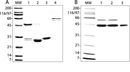
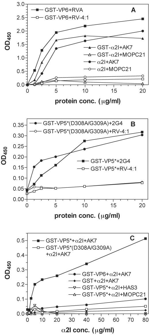
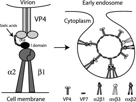
Integrins alpha2beta1, alphaXbeta2, and alphaVbeta3 have been implicated in rotavirus cell attachment and entry. The virus spike protein VP4 contains the alpha2beta1 ligand sequence DGE at amino acid positions 308 to 310, and the outer capsid protein VP7 contains the alphaXbeta2 ligand sequence GPR. To determine the viral proteins and sequences involved and to define the roles of alpha2beta1, alphaXbeta2, and alphaVbeta3, we analyzed the ability of rotaviruses and their reassortants to use these integrins for cell binding and infection and the effect of peptides DGEA and GPRP on these events. Many laboratory-adapted human, monkey, and bovine viruses used integrins, whereas all porcine viruses were integrin independent. The integrin-using rotavirus strains each interacted with all three integrins. Integrin usage related to VP4 serotype independently of sialic acid usage. Analysis of rotavirus reassortants and assays of virus binding and infectivity in integrin-transfected cells showed that VP4 bound alpha2beta1, and VP7 interacted with alphaXbeta2 and alphaVbeta3 at a postbinding stage. DGEA inhibited rotavirus binding to alpha2beta1 and infectivity, whereas GPRP binding to alphaXbeta2 inhibited infectivity but not binding. The truncated VP5* subunit of VP4, expressed as a glutathione S-transferase fusion protein, bound the expressed alpha2 I domain. Alanine mutagenesis of D308 and G309 in VP5* eliminated VP5* binding to the alpha2 I domain. In a novel process, integrin-using viruses bind the alpha2 I domain of alpha2beta1 via DGE in VP4 and interact with alphaXbeta2 (via GPR) and alphaVbeta3 by using VP7 to facilitate cell entry and infection.


Figures

FIG. 1.
FIG. 1.
Binding to MA104 cell α2β1 by laboratory-adapted rotaviruses and their reassortants. Data are for MAb AK7 relative to control MAb MOPC21 at the highest concentration used (20 μg/ml). Viruses were tested with serial twofold MAb dilutions (1.2 to 20 μg/ml) in three to five experiments. MOPC21 (1.2 to 40 μg/ml) treatment gave virus titers that were indistinguishable from titers obtained in the absence of MAb. The percent binding blockade of integrin-dependent viruses was AK7 dose dependent. Integrin-independent viruses showed no blockade at any AK7 concentration tested.
FIG. 2.
FIG. 2.
SA11, RRV, and Wa but not CRW-8 bound α2β1 on K562 cells, virus-α2β1 binding was eliminated by anti-α2 MAb AK7, and SA11 binding was increased after α2β1 activation with anti-β1 MAb 8A2. Titers of infectious virus bound in the presence of MAb MOPC21 were indistinguishable from those of virus bound to cells in the absence of antibody. PBJ, K562-PBJ; α2, α2-K562; α3, α3-K562.
FIG. 3.
FIG. 3.
DGEA but not GPRP inhibited SA11 binding, but both DGEA and GPRP inhibited infection. Virus infectivity inhibition experiments are shown in panel A, and virus binding inhibition data are shown in panel B. conc., concentration.
FIG. 4.
FIG. 4.
DGEA inhibited SA11 binding to α2-K562 but not to K562, PBJ-K562, and α3-K562 cells, whereas GPRP had no effect on SA11 binding to these cells. The titers of virus bound to cells are expressed as a percentage of the titers of virus bound to PBJ-K562 cells in the absence of peptide.
FIG. 5.
FIG. 5.
Gel profiles of Coomassie-stained, expressed proteins. (A) Lanes 1 to 4, GST-α2I, thrombin-cleaved GST-α2I, GST, and GST-VP6, respectively. (B) Lanes 1 to 3, GST-VP5*, GST-VP5*(D308A/G309A), and GST-VP5* after GroEL removal (see Materials and Methods), respectively. MW, molecular size standards.
FIG. 6.
FIG. 6.
Expressed α2 I domain and viral proteins contained functional epitopes (A and B), and α2 I domain binding to GST-VP5* required D308/G309 of VP5* (C). (A and B) Expressed protein (1.25 to 20 μg/ml) on the solid phase reacted with MAbs (10 μg/ml) to functional (closed symbol) or unrelated (matching open symbol) epitopes in EIA. GST (1.25 to 20 μg/ml) reacted with test or control MAbs gave optical densities at 450 nm (OD450) of ≤0.06. (C) Viral GST fusion protein or GST (40 μg/ml) on the solid phase, reacted with α2I (2.5 to 80 μg/ml), was detected with anti-α2I MAb AK7, negative control MAb HAS3 (non-I domain anti-α2), or MOPC21 (10 μg/ml) in EIA. GST fusion protein or GST reacted with GST or BSA (2.5 to 80 μg/ml) gave an OD450 of ≤0.05 by EIA. conc., concentration.
FIG. 7.
FIG. 7.
Model for involvement of α2β1, αXβ2, and αVβ3 in early cellular interactions of integrin-using rotaviruses. Initial cell binding could be via low-affinity binding of VP8* to terminal or subterminal SA (and/or of VP4 to other sugars, such as galactose) on glycolipids or glycoproteins (14, 16, 22, 26), including on integrins such as α2β1. Terminal SA binding is not necessary for α2β1 recognition, but SA binding could promote conformational change in VP4, exposing the VP5* DGE region, or could bring VP4 into closer proximity to the α2 I domain. The VP5* binding to the α2 I domain of the activated, extended form of α2β1 via the type I collagen ligand sequence DGE in VP5* could result in integrin clustering and signal transduction, possibly enhanced by the binding of more than one α2β1 molecule per multimeric VP4 spike. This binding could facilitate VP7 interaction with αXβ2 and αVβ3 (perhaps by further conformational change in VP4), produce further integrin clustering, and lead to endocytosis. There is evidence that integrin-using and integrin-independent rotaviruses can be endocytosed, and activated β1 integrins can enter cells by clathrin-mediated endocytosis (4, 41). Adenovirus recognition of αVβ3 leads to both clathrin-mediated endocytosis and macropinocytosis of virus (37). The ability of heat shock cognate protein 70 to inhibit the infectivity of integrin-using rotaviruses (19) is consistent with usage of the clathrin pathway, as this heat shock protein facilitates vesicle uncoating and clathrin recycling to the cell membrane (25). Alternatively, as caveolin-1 associates with α2β1 and αVβ3 (54) and echovirus 1 binding to α2β1 results in virus internalization through caveolae (36), rotavirus recognition of α2β1 and αVβ3 could lead to rotavirus entry through caveolae. It is also possible that rotavirus could be endocytosed via a clathrin- and caveolin-independent route. The ability of methyl-β-cyclodextrin to inhibit rotavirus infectivity suggests involvement of cell membrane cholesterol during virus-cell entry (21). Although cholesterol was proposed to be involved as a lipid raft component (21), cholesterol depletion also inhibits endocytosis. As has been proposed previously, fusogenic VP5* domains could then selectively permeabilize the endosomal membrane, lowering the [Ca2+] surrounding the virions and leading to virus uncoating (4, 15). This reduction in [Ca2+] also could release bound VP4 and VP7 from integrins, as Ca2+ and other divalent cations are necessary for integrin binding to ligands. A direct entry mechanism for rotavirus (28) appears to be less likely than endocytosis (4, 17) but also would be compatible with integrin usage.

Similar articles

Cited by

References

    1. Barton, E. S., J. C. Forrest, J. L. Connolly, J. D. Chappell, Y. Liu, F. J. Schnell, A. Nusrat, C. A. Parkos, and T. S. Dermody. 2001. Junction adhesion molecule is a receptor for reovirus. Cell 104:441-451. - PubMed
    1. Bergelson, J. M., B. M. Chan, R. W. Finberg, and M. E. Hemler. 1993. The integrin VLA-2 binds echovirus 1 and extracellular matrix ligands by different mechanisms. J. Clin. Investig. 92:232-239. - PMC - PubMed
    1. Bergelson, J. M., N. F. St. John, S. Kawaguchi, R. Pasqualini, F. Berdichevsky, M. E. Hemler, and R. W. Finberg. 1994. The I domain is essential for echovirus 1 interaction with VLA-2. Cell Adhes. Commun. 2:455-464. - PubMed
    1. Chemello, M. E., O. C. Aristimuno, F. Michelangeli, and M. C. Ruiz. 2002. Requirement for vacuolar H+-ATPase activity and Ca2+ gradient during entry of rotavirus into MA104 cells. J. Virol. 76:13083-13087. - PMC - PubMed
    1. Chen, D., J. W. Burns, M. K. Estes, and R. F. Ramig. 1989. Phenotypes of rotavirus reassortants depend upon the recipient genetic background. Proc. Natl. Acad. Sci. USA 86:3743-3747. - PMC - PubMed

Publication types

LinkOut - more resources