. 2003 Sep;77(18):10099-105.
doi: 10.1128/jvi.77.18.10099-10105.2003.

Combinations of two capsid regions controlling canine host range determine canine transferrin receptor binding by canine and feline parvoviruses

Affiliations

Combinations of two capsid regions controlling canine host range determine canine transferrin receptor binding by canine and feline parvoviruses

Karsten Hueffer et al. J Virol. 2003 Sep.

Abstract

Feline panleukopenia virus (FPV) and its host range variant, canine parvovirus (CPV), can bind the feline transferrin receptor (TfR), while only CPV binds to the canine TfR. Introducing two CPV-specific changes into FPV (at VP2 residues 93 and 323) endowed that virus with the canine TfR binding property and allowed canine cell infection, although neither change alone altered either property. In CPV the reciprocal changes of VP2 residue 93 or 323 to the FPV sequences individually resulted in modest reductions in infectivity for canine cells. Changing both residues in CPV to the FPV amino acids blocked the canine cell infection, but that virus was still able to bind the canine TfR at low levels. This shows that both CPV-specific changes control canine TfR binding but that binding is not always sufficient to mediate infection.


Figures
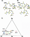
FIG. 1.
FIG. 1.
Structures of CPV and FPV within the areas examined in this study. (A) Atomic model of the structure of the CPV capsid in the vicinity of VP2 residue 93. Labels indicate the VP2 residues that were substituted in these studies. Loop 1 is shown between VP2 residues 91 and 95, while loop 2 is shown between residues 222 and 229. (B) Atomic model of FPV around residue 93 in the same orientation as in panel A. Labels indicate the VP2 residues that were substituted during these studies. Hydrogen bonds between residue 93 and the backbone oxygen atoms of residues 225 and 227 are indicated by green lines. (C) Residues mutated in these studies shown on a roadmap of one asymmetric unit of the CPV capsid (24), showing surface-exposed residues examined in this study. The number following the VP2 residue indicates the relationship of the VP2 molecule containing the residue relative to the reference VP2 monomer in the standard orientation (2×, twofold; 3×, threefold; 5×, fivefold) (1). Residues that were changed in CPV are marked in blue, those changed in FPV are marked in green, and those changed in both viruses are colored in red. Residues 299 and 300, which were not altered in these studies but which also affect canine host range, are shown in brown. Other surface-exposed residues are not shown here. The position of residue 323 in a neighboring asymmetric unit is indicated by the dashed lines.
FIG. 2.
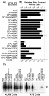
FIG. 2.
Mutants prepared in these studies and their relative titers in feline and canine cells. (A) The genetic background (CPV or FPV) of each virus is indicated, as well as the changes(s) introduced into the VP2 sequence. The single-letter code for amino acids was used, with the letter before the number indicating the amino acid of the wild-type virus and that following indicating the change(s) introduced. (B) Relative infectivity of each virus for canine cells shown as the ratio of the TCID50 titer of the virus stock in A72 cells to that in NLFK cells. Error bars show one standard deviation of the mean for at least three independent experiments. For the CPV-P229A mutant, data from one (ratio, 2.8 log10) of seven independent experiments was excluded from the analysis. (C) Infection and replication in NLFK and A72 cells of selected viruses examined by the production of viral RF and single-stranded DNA (ss). Cells seeded in six-well plates were inoculated with 0.5 TCID50 per cell and were incubated for 2 days, and then the low-molecular-weight DNA was recovered, electrophoresed in an agarose gel, transferred to a membrane, and detected with a 32P-labeled DNA probe. The samples from NLFK cells were exposed for half the exposure of the A72 cell-derived samples.
FIG. 3.
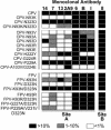
FIG. 3.
Antigenic analysis of the viruses examined by using MAb in an HI assay. Eight hemagglutinin units of virus were incubated for 1 h with twofold dilutions of monoclonal antibodies in the assay, and then erythrocytes were added. The results are shown as the titer of the antibodies for each virus relative to that of wild-type CPV. Filled boxes, mutant HI at 10 to 100% wild-type titer; cross-hatched boxes, less than 10% but greater than 1% of the wild-type titer; open boxes, less than 1% of the wild-type titer.
FIG. 4.
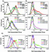
FIG. 4.
Binding of CPV, FPV, or mutant virus capsids to feline or canine cells. Cells were incubated with 10 μg of virus/ml for 1 h at 37°C, and then after cell detachment, fixation, and permeabilization the capsid antigen was detected by antibody staining and flow cytometry. (A) Binding to feline cells of CPV capsids and mutants in a CPV background (upper panel) or FPV capsids and mutants in an FPV background (lower panel). (B) Binding to canine cells of CPV capsid and mutants in a CPV background (upper panel) or FPV capsids and mutants in an FPV background (lower panel). (C) Effect of temperature on binding of CPV or FPV capsids to feline or canine cells. Cells were incubated with 10 μg of virus/ml for 1 h at 4°C or at 37°C, and then after detachment, fixation, and permeabilization viral antigen was detected by antibody staining and flow cytometry.
FIG. 5.
FIG. 5.
Binding of transferrin and capsids of CPV, FPV, or mutant virus to TRVb cells expressing the canine TfR from plasmids. Transiently transfected TRVb cells expressing the canine TfR were incubated with both capsids and Cy5-labeled canine transferrin for 1 h at 37°C, and then after detachment, fixation, and permeabilization the viral antigen was detected by antibody staining and flow cytometry. Virus signal is shown on the X axis, while transferrin is shown on the Y axis. The numbers in the upper quadrants of the graphs represent the percentage of events that fell within those quadrants, and the numbers in parentheses show the percentage of positive cells (as shown by transferrin binding in the upper two quadrants) which bound virus in each case. (A) Binding to the canine TfR expressed on TRVb cells of CPV or mutants derived from CPV. (B) Binding to the canine TfR expressed on TRVb cells as well as on mock-inoculated cells of FPV or mutants derived from FPV.

Similar articles

Cited by

References

    1. Agbandje, M., R. McKenna, M. G. Rossmann, M. L. Strassheim, and C. R. Parrish. 1993. Structure determination of feline panleukopenia virus empty particles. Proteins 16:155-171. - PubMed
    1. Agbandje-McKenna, M., A. L. Llamas-Saiz, F. Wang, P. Tattersall, and M. G. Rossmann. 1998. Functional implications of the structure of the murine parvovirus, minute virus of mice. Structure 6:1369-1381. - PubMed
    1. Ball-Goodrich, L. J., R. D. Moir, and P. Tattersall. 1991. Parvoviral target cell specificity: acquisition of fibrotropism by a mutant of the lymphotropic strain of minute virus of mice involves multiple amino acid substitutions within the capsid. Virology 184:175-186. - PubMed
    1. Ball-Goodrich, L. J., and P. Tattersall. 1992. Two amino acid substitutions within the capsid are coordinately required for acquisition of fibrotropism by the lymphotropic strain of minute virus of mice. J. Virol. 66:3415-3423. - PMC - PubMed
    1. Bloom, M. E., B. D. Berry, W. Wei, S. Perryman, and J. B. Wolfinbarger. 1993. Characterization of chimeric full-length molecular clones of Aleutian mink disease parvovirus (ADV): identification of a determinant governing replication of ADV in cell culture. J. Virol. 67:5976-5988. - PMC - PubMed

Publication types

LinkOut - more resources